[Preprint]. 2025 May 15:2025.05.09.653135.
doi: 10.1101/2025.05.09.653135.

Unreliable homeostatic action potential broadening in cultured dissociated neurons

Affiliations

Unreliable homeostatic action potential broadening in cultured dissociated neurons

Andreas Ritzau-Jost et al. bioRxiv. .

Abstract

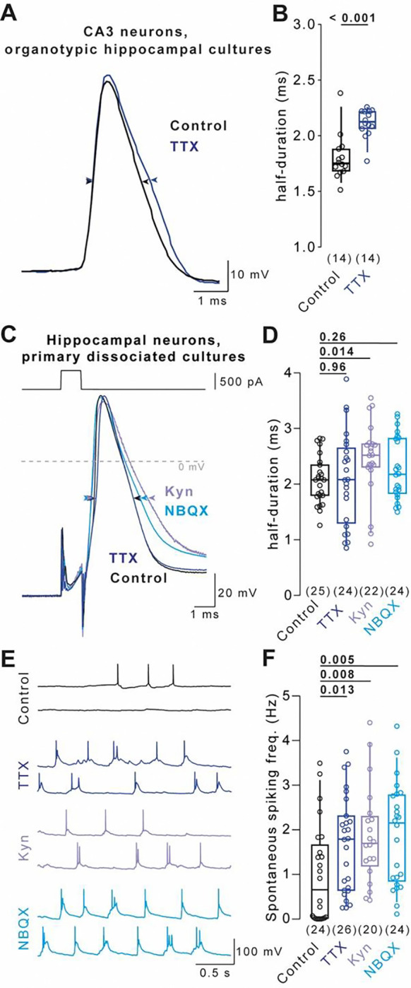
Homeostatic plasticity preserves neuronal activity against perturbations. Recently, somatic action potential broadening was proposed as a key homeostatic adaptation to chronic inactivity in neocortical neurons. Since action potential shape critically controls calcium entry and neuronal function, broadening provides an attractive homeostatic feedback mechanism to regulate activity. Here, we report that chronic inactivity induced by sodium channel block does not broaden action potentials in neocortical neurons under a wide range of conditions. In contrast, action potentials were broadened in CA3 neurons of organotypic hippocampal cultures by chronic sodium channel block and in hippocampal dissociated cultures by chronic synaptic block. Mechanistically, BK-type potassium channels were proposed to underly inactivity-induced action potential broadening. However, BK channels did not affect action potential duration in our recordings. Our results indicate that action potential broadening can occur in specific neurons and conditions but is not a general mechanism of homeostatic plasticity in cultured neurons.


Conflict of interest statement

Declaration of interests The authors declare no competing interests.

Figures

Figure 1
Figure 1. Cell type- and model-specific AP broadening in hippocampal neurons
(A) Example evoked APs recorded from CA3 pyramidal neurons in organotypic hippocampal cultures under control condition (black) and following 48 hours of TTX-treatment (blue). (B) AP durations recorded under conditions in A. (C) Example depolarization-evoked APs (500 pA for 1 ms) recorded from neurons in primary dissociated hippocampal cultures under control condition (black) and following 48 hours of TTX- (blue), Kyn- (purple), or NBQX-treatment (turquoise). (D) AP durations recorded under conditions in C. (E) Example spontaneous APs recorded under the conditions in C. (F) Spontaneous AP frequency under the conditions in C. Numbers in brackets reflect number of recorded neurons. Box plots as median ± interquartile range, whiskers indicate 10–90 percentile. P-values above graphs, significance tested by Mann-Whitney-U test.
Figure 2
Figure 2. Stable AP duration in primary dissociated neurons during TTX-induced homeostatic plasticity
(A) Example current injection-evoked APs (500 pA for 1 ms) under control condition (black) and following 48 hours of TTX-treatment (blue). AP amplitudes are recorded between the membrane potential preceding the AP and AP peak (arrows), AP durations are recorded at half-maximal amplitude (arrow heads). (B) AP duration in control and TTX-treated cells (color code as in A) for eleven different experimental conditions (I–XI). Specification of individual conditions is provided in Table S1. Condition XI adopted from Fig. 1D. (C) Merge of AP durations in control and TTX-treated cells across conditions I–IX normalized to the corresponding condition’s control median duration. (D) AP amplitudes in control and TTX-treated cells as in B. (E) Cross-conditional merge of AP amplitudes in control and TTX-treated cells as in C. (F) Example hyperpolarization-evoked “rebound APs” (−150 pA or −60 pA for 500ms) in control and TTX-treated cells (color code as in A). (G) Rebound AP duration in control and TTX-treated cells as in B. (H) Cross-conditional merge of normalized durations as in C for rebound APs. Numbers in brackets reflect number of recorded neurons. Box plots as median ± interquartile range, whiskers indicate 10–90 percentile. P-values provided above graphs; significance tested using Mann-Whitney-U tests.
Figure 3
Figure 3. Homeostatic plasticity increases network activity and excitatory synaptic transmission
(A) Left: Spontaneously active control (black) and TTX-treated cells (blue) in condition I–IV and VII. Right: Active neurons merged across conditions I–IV and VII. (B) Example spontaneous AP bursts in control and TTX-treated cells. Asterisks mark individual APs. (C) AP number per burst in control and TTX-treated cells across conditions I–IV and VII. (D) Example spontaneous excitatory postsynaptic currents (sEPSCs) in control and TTX-treated cells. (E) Left: sEPSC frequency and Right: sEPSC amplitude in control recordings and after TTX-treatment (merged data from conditions I and II). Numbers in brackets reflect number of recorded neurons. Bars in A reflect fraction of spontaneously active neurons. Whiskers in A reflect confidence limits for 95% confidence interval based on Wilson interval. Box plots as median ± interquartile range, whiskers indicate 10–90 percentile. P-values above graphs, significance tested by Fisher’s exact test in A or Mann-Whitney-U test elsewhere.
Figure 4
Figure 4. Lack of BK channel-dependent AP broadening in dissociated cultured neurons
(A) Left: Example current injection-evoked APs (500 pA for 1 ms) in control solution (black) and solution containing 300 nM Iberiotoxin (IbTx, green). Right: AP duration in control and TTX-treated cells recorded IbTx-free (black and blue, respectively) or IbTx-containing solution (green). Data recorded under condition I. (B) Merged AP duration across control and TTX-treated cells for recordings in IbTx-free and IbTx-containing solution (data in A normalized to the respective group’s median duration in control solution). (C) APs evoked by a 200 ms current injection (top) and overlay of the forst three evoked APs (bottom). (D) Broadening of AP duration of the 3 first APs during 200 ms current injections in control (black, left) and TTX-treated cells (blue, right). Bars as mean ± SEM, lines with markers depict recordings from individual neurons. (E) Left: Example Ace-mNeon-recorded somatic, proximal axonal, and distal axonal APs in a primary hippocampal cultured neuron in IbTx-free solution (black) and following BK channel block by IbTx (100 nM, green). Right: Ratio of somatic and axonal AP durations in IbTx-free and IbTx-containing solution. (F) Left: Example AP train (5 × 20 Hz) and overlay of 1st and 5th train AP at the soma, proximal, and distal axon in control condition (grey and black) and following BK channel block by IbTx (yellow and green). Right: AP broadening (ratio of 5th over 1st train AP duration) in the three compartments in control and IbTx-containing solution. (G) Somatic APs under control condition (grey), following treatment with 4-AP (black), and following treatment with 30μM 4-AP + 100 nM IbTx (green). Numbers in brackets reflect number of recorded neurons. Box plots as median ± interquartile range, whiskers indicate 10–90 percentile. Plots in D–F as mean ± SEM. P-values above graphs, significance tested by Mann-Whitney-U test.
Figure 2 – figure supplement 1
Figure 2 – figure supplement 1. Additional information on recording conditions
The table lists additional relevant information on the experimental conditions I – XI.
Figure 2 – figure supplement 2
Figure 2 – figure supplement 2. Absence of TTX-induced AP broadening in further selected conditions
(A) Box plots of AP durations under control condition (black) or upon TTX-treatment (blue) recorded in condition I either unselected or selected for parameters reflecting unimpaired neuronal function. (B) AP durations as in A for recordings in condition VII. (C) AP durations as in A for recordings in condition VIII. Numbers of recorded neurons provided in brackets. Box plots depict median ± interquartile range, whiskers reflect 10–90 percentile. P-values calculated by Mann-Whitney-U test.
Figure 4 – figure supplement 1
Figure 4 – figure supplement 1. BK channel block does not affect rebound AP duration
(A) Example rebound APs in control solution (black) and solution containing 300 nM Iberiotoxin (IbTx, green). (B) AP duration in control and TTX-treated cells in IbTx-free (black and blue, respectively) or IbTx-containing solution (green). Data recorded in condition I. (C) Merged AP duration across control and TTX-treated cells for recordings in IbTx-free and IbTx-containing solution (data in A, normalized to the respective group’s median duration in IbTx-free solution). Numbers of recorded neurons provided in brackets. Box plots depict median ± interquartile range, whiskers reflect 10–90 percentile. P-values calculated by Mann-Whitney-U test.

References

    1. Borst J.G.G., and Sakmann B. (1999). Effect of changes in action potential shape on calcium currents and transmitter release in a calyx-type synapse of the rat auditory brainstem. Philosophical Transactions of the Royal Society B: Biological Sciences 354, 347–355. 10.1098/RSTB.1999.0386. - DOI - PMC - PubMed
    1. Christie B.R., Eliot L.S., Ito K.I., Miyakawa H., and Johnston D. (1995). Different Ca2+ channels in soma and dendrites of hippocampal pyramidal neurons mediate spike-induced Ca2+ influx. J Neurophysiol 73, 2553–2557. 10.1152/jn.1995.73.6.2553. - DOI - PubMed
    1. Watanabe S., Hoffman D.A., Migliore M., and Johnston D. (2002). Dendritic K+ channels contribute to spike-timing dependent long-term potentiation in hippocampal pyramidal neurons. Proc Natl Acad Sci U S A 99, 8366–8371. 10.1073/PNAS.122210599. - DOI - PMC - PubMed
    1. Bean B.P. (2007). The action potential in mammalian central neurons. Nat Rev Neurosci 8, 451–465. 10.1038/nrn2148. - DOI - PubMed
    1. Debanne D., Campanac E., Bialowas A., Carlier E., and Alcaraz G. (2011). Axon physiology. Physiol Rev 91, 555–602. 10.1152/PHYSREV.00048.2009. - DOI - PubMed

Publication types

LinkOut - more resources