[Preprint]. 2025 May 15:2025.05.13.653829.
doi: 10.1101/2025.05.13.653829.

Cryo-EM evidence for a common factor in Alzheimer's and other neurodegenerations

Affiliations

Cryo-EM evidence for a common factor in Alzheimer's and other neurodegenerations

Leslie R Bridges. bioRxiv. .

Abstract

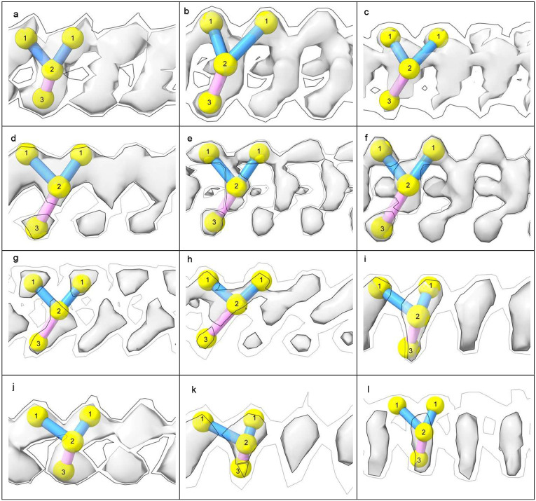
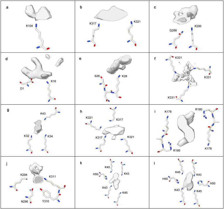
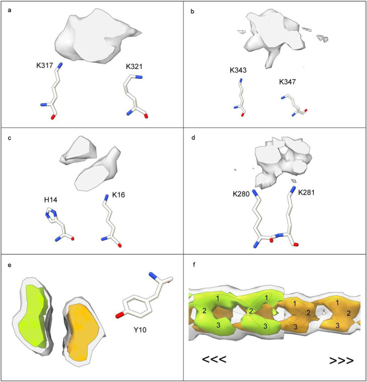
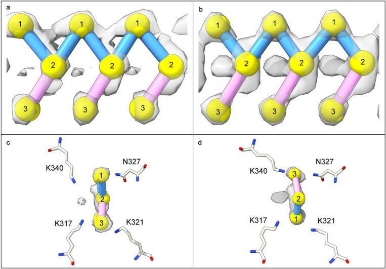
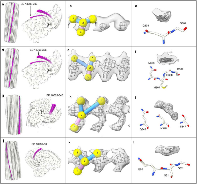
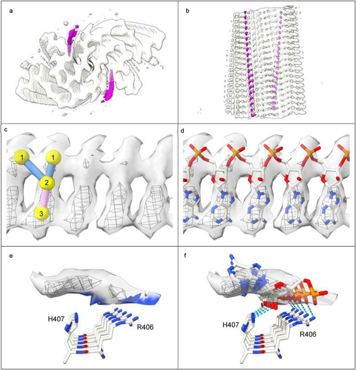
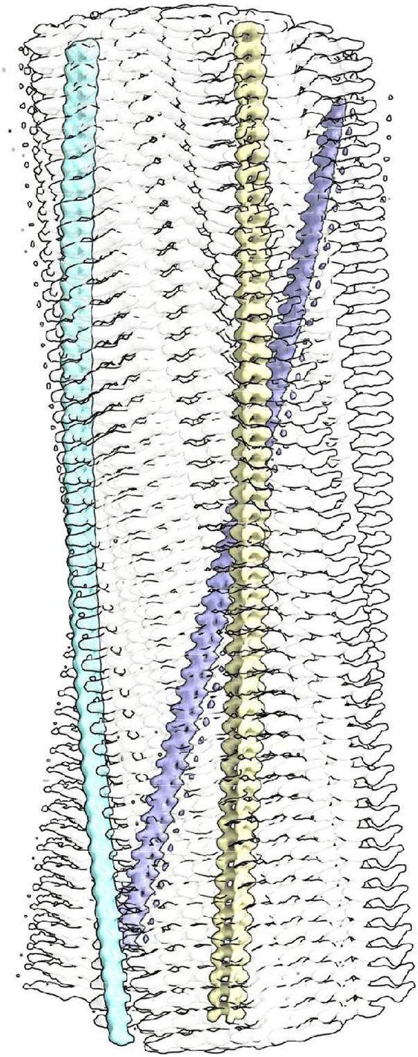
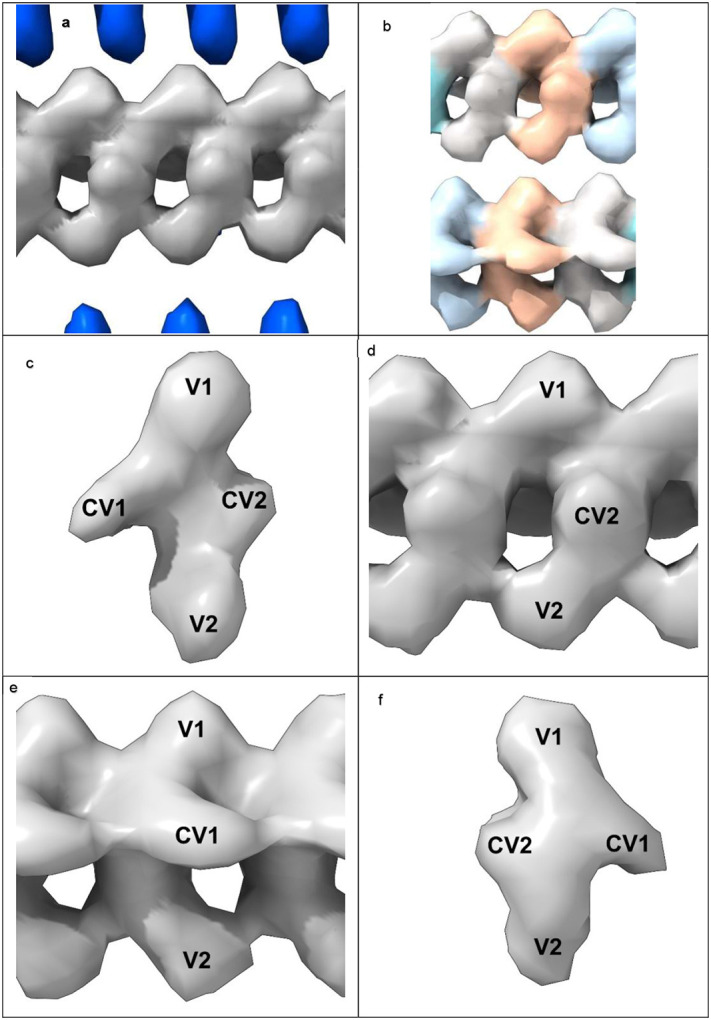
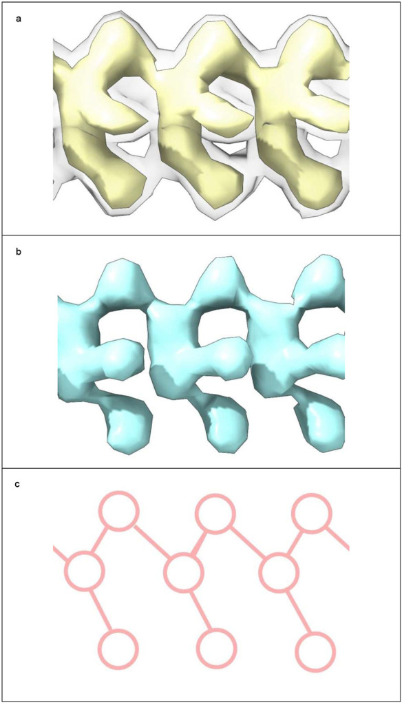
In the last seven years, cryo-EM maps of neuropathological fibrils from Alzheimer's disease and other neurodegenerations have been released by various authors1-44. The first publication11 noted an unknown component coordinating with lysine residues in the protein, a finding recapitulated in many succeeding studies. Previous authors have emphasized difficulties in analysing this component12,20,28,33,43,45, but current findings, using powerful visualisation software UCSF ChimeraX46 on all publicly available maps1-44, indicate that the issue is tractable. Lysine-coordinating extra densities have common features, including a Y-shaped substructure, suggestive of a molecular factor in common, in neuropathological fibrils from a wide range of neurodegenerations and involving misfolded proteins beta-amyloid10,35, alpha-synuclein27,37,39,41, prion protein17, tau1,5,7,8,11,12,15,16,19,22-26,29-33,35,43 and transmembrane protein 106B5,9,18,20,24,28,36,44. A similar component, albeit in non-lysine environments, was found in neuropathological fibrils involving TAR DNA-binding protein 432,3 and TATA-binding protein-associated factor 1536. The results suggest the existence of a common molecular factor, a predominantly anionic polymer, linking these diseases and raising the possibility of a unitary basis for Alzheimer's and other neurodegenerations. Based on evidence here, RNA is a feasible candidate for this putative common factor. Such findings raise the possibility of new diagnostic tests and treatments for these devastating diseases in the future.


Figures

Extended Data Fig. 1 ∣
Extended Data Fig. 1 ∣. LCEDs are a common feature of diverse neuropathological fibrils.
Protein white, LCEDs fuchsia, spacing of protein rungs about 4.8 Å. Images of fibrils created with UCSF ChimeraX. a. Aβ fibril in AD, EMD-15770, b. PrP fibril in GSS, EMD-26607, c. TMEM106B fibril in FTLD-TDP, EMD-26290, d. α-syn fibril in MSA, EMD-10652, e. α-syn fibril in PD, EMD-15285, f. α-syn fibril in JOS, EMD-16189, g. tau fibril in AD, SF, EMD-0260, h. tau fibril in CTE, type II, EMD-0528, i. tau fibril in CBD, EMD-10514, j. tau fibril in PSP, EMD-13218, k. tau fibril in GGT, EMD-13221, l. tau fibril in LNT, EMD-13225.
Extended Data Fig. 2 ∣
Extended Data Fig. 2 ∣. LCEDs are typically Y-shaped.
Shown at two contour levels with markers, authors’ level shown with bold outline, features repeat at about 4.8 Å. Images of fibrils created with UCSF ChimeraX. a. tau fibril in CBD at K290, EMD-10514, b. α-syn fibril in MSA at K43, EMD-10650, c. tau fibril in PART at K317, EMD-12550, d. tau fibril in GGT at K317, EMD-13219, e. tau fibril in LNT at K317, EMD-13224, f. tau fibril in LNT at K317, EMD-13225, g. Aβ fibril in AD at S26, EMD-15770, h. TMEM106B fibril in aFTLD at K178, EMD-18240, i. tau fibril in PSP at K317, EMD-26268, j. PrP fibril in GSS at K104, EMD-26613, k. tau fibril in AD at K311, EMD-26665, l. TMEM106B fibril in MSTD at K139, EMD-28943.
Extended Data Fig. 3 ∣
Extended Data Fig. 3 ∣. LCEDs with an X-shaped substructure are consistent with a Y-shaped constituent molecule.
LCEDs shown at authors’ level with markers. Images of fibrils created with UCSF ChimeraX. a. c. and e. tau fibril in LNT at K317, EMD-13224, PDB 7p6b. b. d. and f. tau fibril in LNT at K317, EMD-13223, PDB 7p6a. In a. and c., viewed from different angles, the LCED is naturally Y-shaped. An X-shape was created artificially in e. by superimposition of the Y-shaped poses in a. and c. In b. d. and f. the LCED is naturally X-shaped, but Y-shaped markers can be fitted either way up, indicating that the X-shape is consistent with averaging of alternative Y-shaped poses.
Extended Data Fig. 4 ∣
Extended Data Fig. 4 ∣. LCEDs occur in stereotyped protein environments.
LCEDs in transverse section opposite 1-6 polar residues. The chemical environments are either purely donor or mixed, donor and acceptor, for hydrogen-bonding purposes. LCEDs shown at authors’ level (light grey) or at two levels (authors’ level darker and opaque). Images of fibrils created with UCSF ChimeraX. a. PrP fibril in GSS at K104, EMD-26613, PDB 7un5, b. tau fibril in AD, PHF, at K317, EMD-15772, PDB 8azu, c. tau fibril in GGT at Q288, EMD-13221, PDB 7p68, d. Aβ fibril in Down’s at D1, EMD-40421, PDB 8sel, e. Aβ fibril in AD at S26, EMD-15770, PDB 8azs, f. tau fibril in AD V337M, triplet filament, at K331, note 3-fold symmetry, EMD-19852, PDB 9eoe, g. α-syn fibril in PD at K32, EMD-15285, PDB 8a9l, h. tau fibril in PART, SF, at K317, EMD-12550, PDB 7nrs, i. TMEM106B fibril in MSTD at K178, note 2-fold symmetry, EMD-28943, PDB 8f9k, j. tau fibril in FTDP-17 P301T at K294, EMD-51320, PDB 9gg1, k. α-syn fibril in MSA at K43, EMD-10650, PDB 6xyo, l. α-syn fibril in MSA at K43, EMD-10652, PDB 6xyq.
Extended Data Fig. 5 ∣
Extended Data Fig. 5 ∣. LCEDs are sometimes bulky.
LCEDs in transverse section opposite protein residues at authors’ level (a-d). Also shown is a bulky non-LCED (e and f). Images of fibrils created with UCSF ChimeraX. a. tau fibril in AD, PHF, at K317, EMD-3742, PDB 5o3o. b. tau fibril in CBD at K343, EMD-10512, PDB 6tjo. c. Aβ fibril in Down’s at H14, note duplex, EMD-40419, PDB 8sek. d. tau fibril in LNT at K280, EMD-13223, PDB 7p6a. e. and f. Aβ fibril in AD/CAA at Y10, note duplex, EMD-18509, PDB 8qn7, authors’ level grey transparent, increased level colours opaque, in transverse section (e) and lengthwise (f). The non-LCED is explicitly divided into two chains. In f. one of the chains is cut away to show the other chain behind. The directions of the two chains are opposite (antiparallel).
Extended Data Fig. 6 ∣
Extended Data Fig. 6 ∣. LCEDs and protein folding.
The spatial relationships of LCEDs are consistent with an effect on protein folding. a. LCED in α-syn fibril of MSA at K43, at two levels, authors’ level opaque, note connections between extra density and protein, EMD-10650. LCEDs (authors’ levels) and protein models from: b. tau fibril of PSP at K317, LCED lies horizontally, EMD-13218 and PDB 7p65, c. tau fibril of GGT at K317, LCED lies vertically, EMD-13219 and PDB 7p66, d. tau fibril of CBD at K290, EMD-10514 and PDB 6tjx, e. tau fibril of AGD at K290, note involvement of E372 (compare to d), EMD-13226 and PDB 7p6d. Images created with UCSF ChimeraX.
Extended Data Fig. 7 ∣
Extended Data Fig. 7 ∣. LCEDs adopt alternative binding poses.
LCEDs shown at authors’ level with markers. Images of fibrils created with UCSF ChimeraX. a. and c. tau fibril in LNT at K317, EMD-13224, PDB 7p6b. b. and d. tau fibril in LNT at K317, EMD-13225, PDB 7p6c. At top, LCEDs are shown lengthwise and at bottom they are shown in cross-section, in their protein environments. Although the morphologies are virtually identical, the binding poses are opposite, as demonstrated by the position of the markers relative to the coordinating residues in the lower frames.
Extended Data Fig. 8 ∣
Extended Data Fig. 8 ∣. Rod-like densities also occur in non-lysine environments.
Note similarities with LCEDs, including rod-like structure (a, d, g and j, protein white, non-LCEDs fuchsia, spacing of protein rungs about 4.8 Å), Y-shaped substructure (b, e, h and k, at two levels with markers, authors’ level in mesh, features repeat at about 4.8 Å) and proximity to proton donor and acceptor atoms in the protein (c, f, i and l, at two levels, author’s level in mesh, opposite coordinating residues). Images of fibrils created with UCSF ChimeraX a. b. and c. TDP-43 fibril in ALS-FTLD at Q303, EMD-13708, PDB 7py2 d. e. and f. TDP-43 fibril in ALS-FTLD at N306, EMD-13708, PDB 7py2 g. h. and i. TDP-43 fibril in FTLD-TDP type A at Q343, EMD-16628, PDB 8cg3. j. k. and l. TAF15 fibril in aFTLD at Q60, EMD-16999, PDB 8ons.
Extended Data Fig. 9 ∣
Extended Data Fig. 9 ∣. Rod-like densities in RNA-induced tau fibrils are similar to LCEDs.
a. and b. Protein white, rod-like extra densities (containing RNA) fuchsia, spacing of protein rungs about 4.8 Å. c. and d. Extra density at two contour levels, authors’ level in mesh, with markers (c) and authors’ model of RNA (d). e. and f. Extra density in transverse section opposite H407 and R406 basic polar residues, at two levels, authors’ level in mesh. e. shows colour zone (blue shading) at 3.5 Å from donor atoms in the protein. f. shows authors’ model of RNA forming hydrogen bonds with donor atoms in histidines and arginines (dashed blue lines). Images of EMD-25364 and PDB 7sp1 created with UCSF ChimeraX.
Fig. 1 ∣
Fig. 1 ∣. Lysine coordinating extra densities are like supporting rods.
Alpha-synuclein fibril of multiple system atrophy. LCEDs (colours) are upright, parallel to the fibril and at right angles to protein rungs (transparent, white), consistent with a structural role as guide and support. At authors’ contour level. Distance between protein rungs is 4.7 Å. Image of EMD-10650 created with UCSF ChimeraX.
Fig. 2 ∣
Fig. 2 ∣. LCEDs are consistent with an underlying polymer.
LCED at K43 in α-syn fibril of MSA, at authors’ contour level. a. LCED (grey) is at right angles to the protein (blue rungs). b. Repeat distance 4.7 Å. c.-f. Elliptic rod with distinct features at vertices, V1 and V2, and co-vertices, CV1 and CV2. The well-defined repeating features, continuous and in step with protein, are consistent with an underlying polymer. Images of EMD-10650 created with UCSF ChimeraX. Colour-shading of repeating units in b. by Segger.
Fig. 3 ∣
Fig. 3 ∣. LCEDs are consistent with a molecular factor in common.
a. LCED from α-syn fibril of MSA, at two contour levels (authors’ level transparent grey). b. tau fibril of LNT, at authors’ level. The LCEDs, involving completely different proteins and diseases, have common features and Y-shaped connectivity, consistent with a constituent molecule in common, a polymer. Images of EMD-10650 and EMD-13225 created with UCSF ChimeraX. Connectivity diagram (c) created with ImageJ.

Similar articles

References

    1. Arakhamia T. et al. Posttranslational modifications mediate the structural diversity of tauopathy strains. Cell 180, 633–644.e12 (2020). - PMC - PubMed
    1. Arseni D. et al. Structure of pathological TDP-43 filaments from ALS with FTLD. Nature 601, 139–143 (2022). - PMC - PubMed
    1. Arseni D. et al. TDP-43 forms amyloid filaments with a distinct fold in type A FTLD-TDP. Nature 620, 898–903 (2023). - PMC - PubMed
    1. Arseni D. et al. Heteromeric amyloid filaments of ANXA11 and TDP-43 in FTLD-TDP type C. Nature 634, 662–668 (2024). - PMC - PubMed
    1. Chang A. et al. Homotypic fibrillization of TMEM106B across diverse neurodegenerative diseases. Cell 185, 1346–1355.e15 (2022). - PMC - PubMed

Publication types

LinkOut - more resources