[Preprint]. 2025 May 18:2025.05.16.654332.
doi: 10.1101/2025.05.16.654332.

Embryonic Stem Cell-Specific Responses to DNA Replication Stress

Affiliations

Embryonic Stem Cell-Specific Responses to DNA Replication Stress

Ryan C James et al. bioRxiv. .

Abstract

Genome maintenance is of the utmost importance in stem cells, as mutations can be propagated and cause defects in derivative tissues. Many stem cell types display low mutation rates, with embryonic stem cells (ESCs) being a notable example. The bases for this property are unclear but may be achieved by optimization of various processes including high-fidelity DNA repair, cell cycle checkpoint controls, and hypersensitivity to genotoxic insults that trigger cell death. Here, we investigate the mechanisms underlying the unique responses of mouse ESCs (mESCs) to replication stress (RS) using an array of small molecule inhibitors and genotoxins. We find that whereas mESCs survive under acute RS in an ATR- and CHK1-dependent manner similar to somatic cells, they lack a strong G2/M checkpoint and fail to repair DNA crosslinks in the absence of ATR signaling. Despite the lack of a strong G2/M checkpoint, mESCs maintain a spindle assembly checkpoint (SAC). We posit that mESCs preferentially repair DNA crosslinks in S phase via homology-directed mechanisms, and cells that fail to complete repair before mitosis undergo mitotic catastrophe and cell death. These findings shed light on mutation avoidance mechanisms in ESCs that may extend to other stem cell types.


Figures

Figure 1.
Figure 1.. Mouse ESC and MEF responses to agents that induce replication stress.
(A) Western blots for p-CHK1 (S345), CHK1, p-CDK1 (Thr14, Tyr15) of mESCs or MEFs (B) treated with indicated drugs for 24 hours. (C) DNA content analysis of mESCs or MEFs (D) treated with the indicated drugs for 24 hours. Veh, vehicle; Bleo, bleomycin; MMC, mitomycin C; MELP, melphalan; APH, aphidicolin.
Figure 2.
Figure 2.. Survival of ESCs under acute replication stress is CHK1-dependent.
(A) DNA content analysis of mESCs or (B) MEFs treated with the indicated drugs for 24 hours. Drugs were added simultaneously. (C) Viability analysis via MTT assay of mESCs or (D) MEFs. MTT intensity is normalized to an untreated sample. Error bars show SD.
Figure 3.
Figure 3.. The response of mESCs to crosslinks Is not CHK1-dependent.
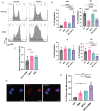
(A) Representative images of γH2A.X staining of mESCs treated with MMC for 24 hours. (B) Quantification of γH2A.X foci per nuclear slice. Z=3, n>150, N=3. (C) DNA content analysis of mESCs treated with the indicated drugs for 24 hours. Drugs were added simultaneously. (D) Quantification of C. Error bars show SD. (E) Hallmark enrichment plot (MMC-treated vs Vehicle) of the top and bottom 10 hallmarks from RNA sequencing of mESCs treated with MMC for 24 hours.
Figure 4.
Figure 4.. ESCs rely on the SAC to prevent chromosomal abnormalities in response to replication stress.
(A) DNA content analysis of mESCs treated with the indicated drugs for 24 hours. Drugs were added simultaneously. (B) Quantification of A. Error Bars show SD. (C) Viability analysis via MTT assay of mESCs treated with the indicated drugs. (D) Representative image of micronucleus formed during treatment with MPS1i+MMC. (E) Quantification of D shows the %of pairs of cells with a micronucleus between them. 3 biological replicates with 20 pairs analyzed each.
Figure 5.
Figure 5.. Differential responses of ESCs and MEFs to the inhibition of ATR and ATM.
(A) Viability analysis via MTT of mESCs or MEFs (B) treated with the indicated drugs. (C) Flow cytometry analysis of mESCs treated with indicated drugs. (D) Quantification of C. (E) Flow cytometry analysis of MEFs treated with indicated drugs. (F) Quantification of C. All error bars show SD

Similar articles

References

    1. Mulas C., Kalkan T., von Meyenn F., Leitch H.G., Nichols J., and Smith A. (2019). Defined conditions for propagation and manipulation of mouse embryonic stem cells. Development 146. 10.1242/dev.173146. - DOI - PMC - PubMed
    1. Ballabeni A., Park I.-H., Zhao R., Wang W., Lerou P.H., Daley G.Q., and Kirschner M.W. (2011). Cell cycle adaptations of embryonic stem cells. Proc Natl Acad Sci USA 108, 19252–19257. 10.1073/pnas.1116794108. - DOI - PMC - PubMed
    1. Coronado D., Godet M., Bourillot P.-Y., Tapponnier Y., Bernat A., Petit M., Afanassieff M., Markossian S., Malashicheva A., Iacone R., et al. (2013). A short G1 phase is an intrinsic determinant of naïve embryonic stem cell pluripotency. Stem Cell Res. 10, 118–131. 10.1016/j.scr.2012.10.004. - DOI - PubMed
    1. Neganova I., Tilgner K., Buskin A., Paraskevopoulou I., Atkinson S.P., Peberdy D., Passos J.F., and Lako M. (2014). CDK1 plays an important role in the maintenance of pluripotency and genomic stability in human pluripotent stem cells. Cell Death Dis. 5, e1508. 10.1038/cddis.2014.464. - DOI - PMC - PubMed
    1. Koledova Z., Kafkova L.R., Calabkova L., Krystof V., Dolezel P., and Divoky V. (2010). Cdk2 inhibition prolongs G1 phase progression in mouse embryonic stem cells. Stem Cells Dev. 19, 181–194. 10.1089/scd.2009.0065. - DOI - PubMed

Publication types

LinkOut - more resources