. 2025 May 19:2025:10.17912/micropub.biology.001608.
doi: 10.17912/micropub.biology.001608. eCollection 2025.

Impact of embryo size on apoptosis in C. elegans

Affiliations

Impact of embryo size on apoptosis in C. elegans

Madiha Javeed Ghani et al. MicroPubl Biol. .

Abstract

During C. elegans development 131 somatic cells reproducibly undergo programmed cell death. Many of these 131 cells 'programmed to die' are the smaller daughter of a neuroblast that divides asymmetrically and die through apoptosis. To determine whether cell size impacts the ability of cells programmed to die to undergo apoptosis, we increased or decreased embryo size by RNA interference-mediated knock-down of the genes C27D9.1 or ima-3 , respectively. We found that in apoptosis-compromised genetic backgrounds, C27D9.1 ( RNAi ) enhances and ima-3 ( RNAi ) partially suppresses inappropriate survival of cells programmed to die. This supports the notion that in C. elegans embryos, an increase in cell size compromises and a decrease in cell size promotes the ability of cells programmed to die to undergo apoptosis.


Conflict of interest statement

The authors declare that there are no conflicts of interest present.

Figures
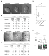
Figure 1.
<b>RNAi-mediated embryo size alterations and impact on the apoptotic death of the NSM sister cell</b>
Figure 1. RNAi-mediated embryo size alterations and impact on the apoptotic death of the NSM sister cell
(A) RNAi-mediated embryo size alterations. DIC images of representative embryos following sorb-1 (RNAi) , ima-3 (RNAi) or C27D9.1 (RNAi) are shown. Scale bars: 10μm. All embryos were analysed at a similar stage of embryonic development (about 220-cell stage). (B) Average embryo length after RNAi-mediated embryo size alterations. For statistical comparison, the lengths of ima-3 (RNAi) or C27D9.1 (RNAi) embryos were compared to the lengths of sorb-1 (RNAi) embryos . Statistical significance was determined by least square means method. p <0.001(***). (C) RNAi-mediated NSM neuroblast (NSMnb) size alterations. Fluorescence images of representative NSMnb following sorb-1 (RNAi) , ima-3 (RNAi) or C27D9.1 (RNAi) are shown. The P pie-1 mCherry::ph PLCΔ reporter ( ltIs44 ) was used to visualize cell boundaries. Scale bars: 3μm. (D) Average NSMnb size measurements after RNAi-mediated embryo size alterations. Each dot represents the estimated size of one NSMnb. The average size for each RNAi treatment is shown above the graph. Statistical significance was determined by Mann-Whitney U-tests. p <0.0001(****). (E) Schematics of the NSM lineage and the NSMsc survival assay. In wild type ( +/+ ), the NSMsc dies and its larger sister differentiates into the NSM neuron, which can be visualized in the anterior pharynx of L3/L4 larvae using the P tph-1 his-24 ::gfp reporter ( bcIs66 ). There are two bilaterally symmetric NSM lineages. For this reason, in +/+ there are two GFP-positive cells in the anterior pharynx. If the NSMsc inappropriately survives ('Mutant'), three to four GFP-positive cells are observed. (F) Average NSMsc survival (%) after RNAi-mediated embryo size alterations. NSM survival for each RNAi treatment was scored in a wild-type ( +/+ ) background and in the background of different ced-3 loss-of-function mutations ( n2438 , n2427 , n2436 , n717 ). All strains used were homozygous for the P tph-1 his-24 ::gfp reporter ( bcIs66 ). For statistical comparison, NSMsc survival in ima-3 (RNAi) or C27D9.1 (RNAi) was compared to NSMsc survival in sorb-1 (RNAi) for each genotype. One sided Fisher's exact tests were used to determine statistical significance. p <0.05 (*), p <0.01(**) and p <0.001(***).

References

    1. Audhya Anjon, Hyndman Francie, McLeod Ian X., Maddox Amy S., Yates John R., Desai Arshad, Oegema Karen. A complex containing the Sm protein CAR-1 and the RNA helicase CGH-1 is required for embryonic cytokinesis in Caenorhabditis elegans . The Journal of Cell Biology. 2005 Oct 24;171(2):267–279. doi: 10.1083/jcb.200506124. - DOI - PMC - PubMed
    1. Bellanger Jean-Michel, Gönczy Pierre. TAC-1 and ZYG-9 Form a Complex that Promotes Microtubule Assembly in C. elegans Embryos. Current Biology. 2003 Sep 1;13(17):1488–1498. doi: 10.1016/s0960-9822(03)00582-7. - DOI - PubMed
    1. Benjamini Yoav, Hochberg Yosef. Controlling the False Discovery Rate: A Practical and Powerful Approach to Multiple Testing. Journal of the Royal Statistical Society Series B: Statistical Methodology. 1995 Jan 1;57(1):289–300. doi: 10.1111/j.2517-6161.1995.tb02031.x. - DOI
    1. Brenner S. THE GENETICS OF CAENORHABDITIS ELEGANS . Genetics. 1974 May 1;77(1):71–94. doi: 10.1093/genetics/77.1.71. - DOI - PMC - PubMed
    1. Burton Nick O., Burkhart Kirk B., Kennedy Scott. Nuclear RNAi maintains heritable gene silencing in Caenorhabditis elegans . Proceedings of the National Academy of Sciences. 2011 Nov 21;108(49):19683–19688. doi: 10.1073/pnas.1113310108. - DOI - PMC - PubMed

LinkOut - more resources