

This is a preprint.
GLAPAL-H: Global, Local, And Parts Aware Learner for Hydrocephalus Infection Diagnosis in Low-Field MRI
- PMID: 40463531
- PMCID: PMC12132119
- DOI: 10.1101/2025.05.14.25327461
GLAPAL-H: Global, Local, And Parts Aware Learner for Hydrocephalus Infection Diagnosis in Low-Field MRI
Update in
-
GLAPAL-H: Global, Local, And Parts Aware Learner for Hydrocephalus Infection Diagnosis in Low-Field MRI.IEEE Trans Biomed Eng. 2025 Jun 9;PP:10.1109/TBME.2025.3578541. doi: 10.1109/TBME.2025.3578541. Online ahead of print. IEEE Trans Biomed Eng. 2025. PMID: 40489263
Abstract
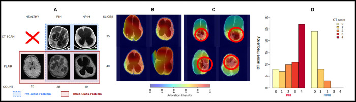
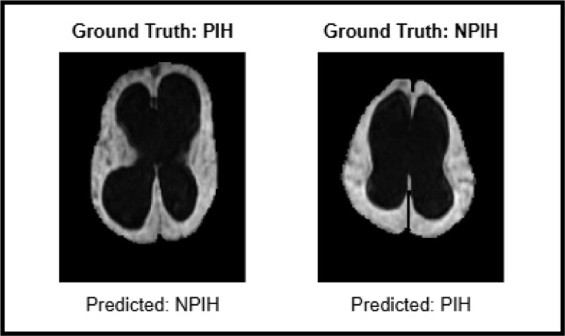
Objective: The study aims to develop a method for differentiating between healthy, post-infectious hydrocephalus (PIH), and non-post-infectious hydrocephalus (NPIH) in infants using low-field MRI, which is a safer, low-cost alternative to CT scans. The study develops a custom approach that captures hydrocephalic etiology while simultaneously addressing quality issues encountered in low-field MRI.
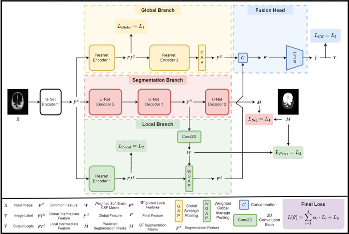
Methods: Specifically, we propose GLAPAL-H, a Global, Local, And Parts Aware Learner, which develops a multi-task architecture with global, local, and parts segmentation branches. The architecture segments images into brain tissue and CSF while using a shallow CNN for local feature extraction and develops a parallel deep CNN branch for global feature extraction. Three regularized training loss functions are developed - one for each of global, local, and parts components. The global regularizer captures holistic features, the local focuses on fine details, and the parts regularizer learns soft segmentation masks that enable local features to capture hydrocephalic etiology.
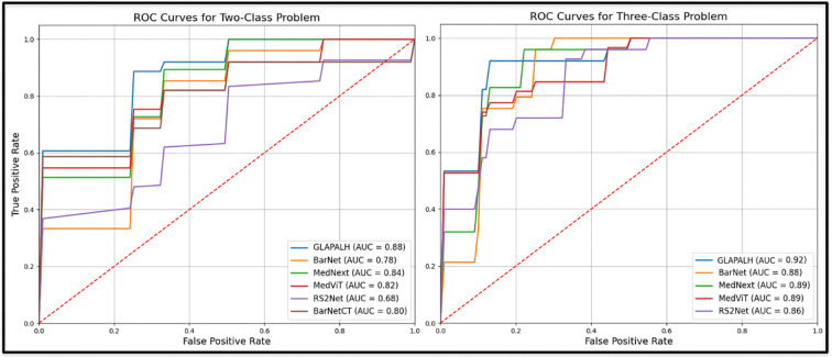
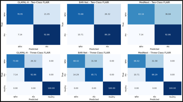
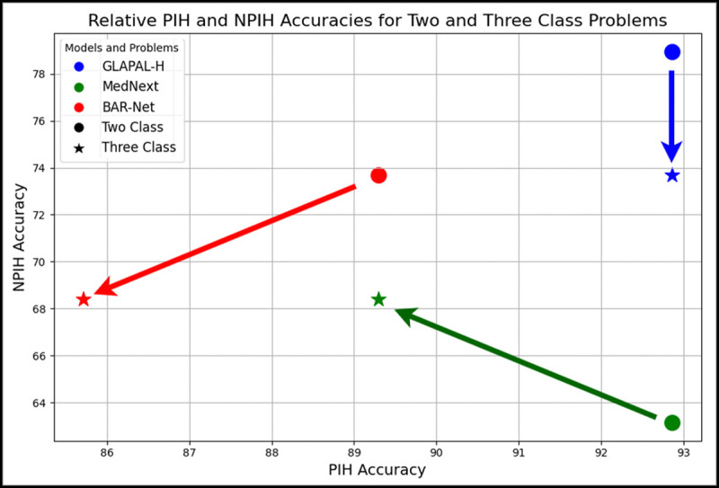
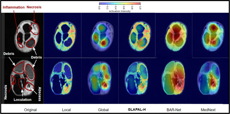
Results: The study's results show that GLAPAL-H outperforms state-of-the-art alternatives, including CT-based approaches, for both Two-Class (PIH vs. NPIH) and Three-Class (PIH vs. NPIH vs. Healthy) classification tasks in accuracy, interpretability, and generalizability.
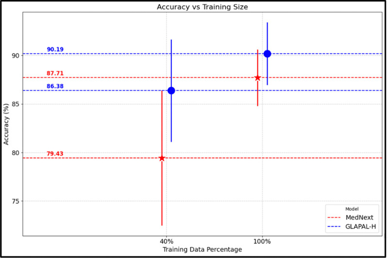
Conclusion/significance: GLAPAL-H highlights the potential of low-field MRI as a safer, low-cost alternative to CT imaging for pediatric hydrocephalus infection diagnosis and management. Practically, GLAPAL-H demonstrates robustness against quantity and quality of training imagery, enhancing its deployability. The code for this work is available here: https://github.com/mukherjeesrijit/glapalh.
Figures
References
-
- Warf B. C.. “Comparison of endoscopic third ventriculostomy alone and combined with choroid plexus cauterization in infants younger than 1 year of age: a prospective study in 550 African children”. J. Neurosurg.: Pediatr. 2005. - PubMed
-
- Dewan M. C. et al. “Global hydrocephalus epidemiology and incidence: systematic review and meta-analysis”. Journal of neurosurgery. 2018. - PubMed
Publication types
Grants and funding
LinkOut - more resources
Full Text Sources