. 2025 Jan-Mar;35(1):79-90.
doi: 10.4103/jcecho.jcecho_15_25. Epub 2025 Apr 30.

How to Do Echo for Noninvasive Hemodynamic Evaluation of the Patient in the Intensive Care Unit: A Consensus Statement of the Italian Society of Echocardiography and Cardiovascular Imaging

Affiliations

How to Do Echo for Noninvasive Hemodynamic Evaluation of the Patient in the Intensive Care Unit: A Consensus Statement of the Italian Society of Echocardiography and Cardiovascular Imaging

Maurizio Cusmà Piccione et al. J Cardiovasc Echogr. 2025 Jan-Mar.

Abstract

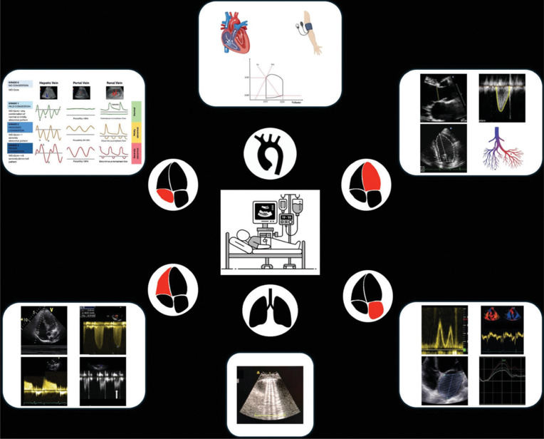
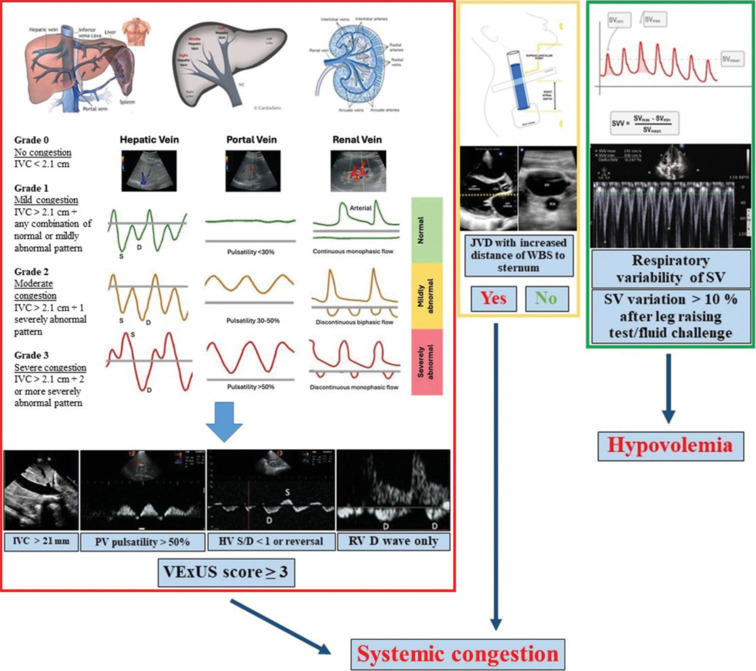
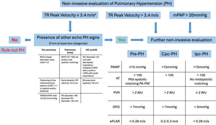
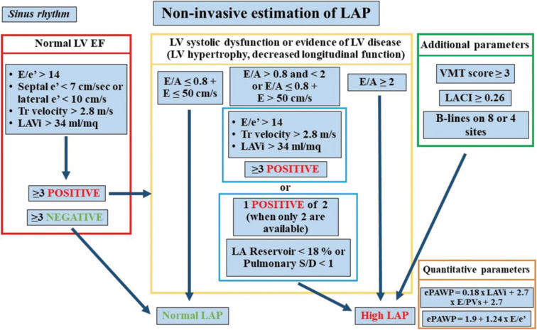
Critically ill patients in the intensive care unit (ICU) require continuous hemodynamic monitoring to guide therapeutic decisions and prevent clinical deterioration. Echocardiography has emerged as a cornerstone for noninvasive hemodynamic assessment, offering real-time, bedside evaluation of key parameters such as venous congestion, pulmonary pressures, left atrial pressure (LAP), systemic vascular resistances, cardiac output, and ventricular-arterial coupling. Systemic venous congestion and right atrial pressure (RAP) can be assessed through inferior vena cava diameter measurement and respiratory variation, with additional accuracy provided by the VeXUS score, which incorporates hepatic, portal, and renal vein Doppler profiles. Internal jugular vein assessment and left ventricular (LV) stroke volume variability further refine RAP estimation. Pulmonary hypertension (PH) and right ventricular dysfunction can be evaluated through echocardiographic markers that differentiate precapillary from postcapillary PH, enabling tailored treatment strategies. In addition, echocardiography is fundamental for detecting right ventricular failure, particularly in PH and cardiogenic shock. LAP and systemic hemodynamics are integral to assessing LV diastolic and systolic dysfunction, which are pivotal in heart failure and cardiogenic shock management. Echocardiography also provides insights into vascular system properties and their interaction with cardiac performance, while lung ultrasound aids in detecting interstitial edema of cardiac origin. As a fast, reliable, and reproducible tool, echocardiography is the gold standard for noninvasive hemodynamic assessment in ICU patients, facilitating prompt and precise therapeutic decisions.

Keywords: Echocardiography; hemodynamic monitoring; intensive care unit; left atrial pressure; noninvasive monitoring; pulmonary hypertension; right ventricular function; venous congestion.


Conflict of interest statement

There are no conflicts of interest.

Figures

Central Illustration
Stepwise echocardiographic assessment of hemodynamics at the bedside. Key parameters include venous congestion, systemic and pulmonary pressures, left atrial pressure, cardiac output, and lung ultrasound findings, following a progression from simple to advanced measurements to guide rapid clinical decision-making
Figure 1
Figure 1
Red box showing, in the top panel, the parameters needed to calculate VeXUS score, and, in the low panel, severely abnormal pattern of each parameter suggestive of severe congestion. Yellow box showing the algorithm for the estimation of central venous pressure by evaluating the internal jugular vein. Green box showing stroke volume variability as a sign of hypovolemia. HVen: Hepatic vein; IVC: Inferior vena cava; JVD: jugular vein distension; PVen: Portal vein; RAP: Right atrial pressure; RVen: Renal vein; SV: Stroke volume
Figure 2
Figure 2
Noninvasive evaluation of pulmonary hypertension. Starting from the evaluation of tricuspid regurgitation peak velocity or mean pulmonary artery pressure, this approach allows for a noninvasive assessment based on echocardiographic parameters to distinguish between precapillary, postcapillary, or combined pulmonary hypertension. AR: Aortic root; Cpc-PH: Combined post- and precapillary pulmonary hypertension; DPG: Diastolic pressure gradient; ePLAR: Echocardiographic pulmonary-to-left atrial ratio; Ipc-PH: Isolated postcapillary pulmonary hypertension; IVC: Inferior vena cava; LVEI: Left ventricle eccentricity index; LV: Left ventricle; mPAP: Mean pulmonary artery pressure; PA: Pulmonary artery; PA-PW: Pulmonary artery pulsed-Doppler wave; PAWP: Pulmonary arterial wedge pressure; PR: Pulmonary regurgitation; Pre-PH: Precapillary pulmonary hypertension; PVR: Pulmonary vascular resistance; RA: Right atrium; RV: Right ventricle; RVOT AT: Right ventricle outflow tract acceleration time; sPAP: Systolic pulmonary arterial pressure; TAPSE: Tricuspid annular plane systolic excursion; TR: Tricuspid regurgitation; WU: Wood units. aOr unmeasurable
Figure 3
Figure 3
Red box showing the algorithm, in sinus rhythm, for the evaluation of left ventricle (LV) diastolic function in patients with normal LV ejection fraction and without additional signs of LV disease. Yellow box showing the algorithm for the evaluation of LV diastolic function in patients with LV systolic dysfunction and/or pathologic remodeling. Green box showing additional parameters for the estimation of left atrial pressure (LAP). Orange box showing parameters to, numerically, quantify LAP and pulmonary artery wedge pressure. E/A: ratio between peak velocities of mitral E and A; E/e’ ratio between mitral E velocity and mean e’ on tissue Doppler imaging; ePAWP: Estimated pulmonary artery wedge pressure; LACI: Left atrial coupling index; LAP: Left atrial pressure; LA Reservoir, two-dimensional strain of atrial walls during reservoir phase; LAVi: Left atrial volume index; Tr: Tricuspid regurgitation; VMT: Visually assessed time difference between mitral valve and tricuspid valve opening
Figure 4
Figure 4
Example of left atrial pressure estimation in patients with depressed ejection fraction and left ventricle disease, when E/A is between 0.8 and 2 and only two criteria are available (average E/e’, tricuspid regurgitation velocity, and left atrial volume index). In cases where one criterion is positive and another is negative, left atrial reservoir strain can help distinguish between Grade 1 and Grade 2 diastolic dysfunction. In addition, two alternative methods for estimating filling pressures are illustrated: the left atrioventricular coupling index and the visually assessed time difference between mitral valve and tricuspid valve opening (VMT), combined with inferior vena cava size. IVC: Inferior vena cava; LA: Left atria; LACI: Left atrioventricular coupling index; LAP: Left atrial pressure; LAVi: Left atrial volume index; VMT: Visually assessed time difference between mitral valve and tricuspid valve opening
Figure 5
Figure 5
Red box showing the algorithm, in atrial fibrillation, for the evaluation of left atrial pressure (LAP), by using step 1 composed of four parameters, such as mitral E peak velocity, septal E/e’, mitral E deceleration time, Tr velocity, followed, when needed, by a step 2, composed of three parameters, such as pulmonary S/D, body mass index and left atria (LA) Reservoir strain. Green box showing a stepwise algorithm, in atrial fibrillation, for the estimation of LAP, composed of septal E/e’, left atrial volume index, and LA Reservoir strain. BMI: Body mass index; E/e’ ratio between mitral E velocity and septal e’ on tissue Doppler imaging; LAP: Left atrial pressure; LA Reservoir, two-dimensional strain of atrial walls during reservoir phase; LAVi: Left atrial volume index; Pulmonary S/D, ratio between peak velocities Tr, tricuspid regurgitation
Figure 6
Figure 6
Critical care ultrasound in shock. The proposed algorithm allows the classification of different types of shock based on echocardiographic parameters. CI: Cardiac index; LAP: Left atrial pressure; MAP: Mean arterial pressure; RAP: Right atrial pressure; SV: Stroke volume; SVR: Systemic vascular resistances; VAC: Ventricular–arterial coupling; aafter leg raising test/fluid challenge

Similar articles

Cited by

References

    1. Rudski LG, Lai WW, Afilalo J, Hua L, Handschumacher MD, Chandrasekaran K, et al. Guidelines for the echocardiographic assessment of the right heart in adults: A report from the American Society of Echocardiography endorsed by the European Association of Echocardiography, a registered branch of the European Society of Cardiology, and the Canadian Society of Echocardiography. J Am Soc Echocardiogr. 2010;23:685–713. - PubMed
    1. Hussein MF, Mohammad WJ, Essa SO. Echocardiographic evaluation of central venous pressure using inferior vena cava characteristics: An estimate guide for right atrial pressure in intensive care unit. J Cardiovasc Echogr. 2024;34:206–13. - PMC - PubMed
    1. Soliman-Aboumarie H, Denault AY. How to assess systemic venous congestion with point of care ultrasound. Eur Heart J Cardiovasc Imaging. 2023;24:177–80. - PubMed
    1. Istrail L, Kiernan J, Stepanova M. A novel method for estimating right atrial pressure with point-of-care ultrasound. J Am Soc Echocardiogr. 2023;36:278–83. - PubMed
    1. Wang J, Zhou D, Gao Y, Wu Z, Wang X, Lv C. Effect of VTILVOT variation rate on the assessment of fluid responsiveness in septic shock patients. Medicine (Baltimore) 2020;99:e22702. - PMC - PubMed

LinkOut - more resources