Review
. 2025 Jan-Mar;35(1):8-18.
doi: 10.4103/jcecho.jcecho_26_25. Epub 2025 Apr 30.

The Role of Imaging in Cardiovascular Prevention: A Comprehensive Review

Affiliations
Review

The Role of Imaging in Cardiovascular Prevention: A Comprehensive Review

Maria Ludovica Carerj et al. J Cardiovasc Echogr. 2025 Jan-Mar.

Abstract

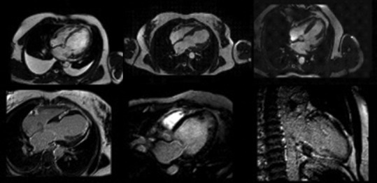
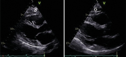
Cardiovascular diseases (CVDs) remain the leading cause of morbidity and mortality worldwide, and traditional preventive measures focus on lifestyle modifications, pharmacologic interventions, and risk stratification. Recently, imaging has emerged as an interesting tool in cardiovascular prevention. This review explores the role of various imaging modalities in early detection, risk assessment, and disease monitoring. Noninvasive techniques such as carotid ultrasound, arterial stiffness assessment, echocardiography, and coronary artery calcium scoring enable the identification of subclinical atherosclerosis and ventricular dysfunction, providing insights that complement conventional risk factors. Coronary computed tomography angiography and cardiac magnetic resonance offer high-resolution visualization of vascular and myocardial pathology, contributing to refined risk stratification. Furthermore, emerging markers such as epicardial adipose tissue and hepatic steatosis are gaining recognition as potential predictors of cardiovascular risk. Advancements in artificial intelligence (AI) are revolutionizing cardiovascular imaging by enhancing image interpretation, automating risk prediction, and facilitating personalized medicine. Future research should focus on optimizing the integration of imaging into clinical workflows, improving risk prediction models, and exploring AI-driven innovations. By exploiting imaging technologies, clinicians could enhance primary and secondary prevention strategies, ultimately reducing the global burden of CVDs.

Keywords: Artificial intelligence; cardiac magnetic resonance; cardiovascular imaging; cardiovascular prevention; echocardiography; epicardial adipose tissue.


Conflict of interest statement

There are no conflicts of interest.

Figures

Figure 1
Figure 1
Mild carotid intima-media thickness. Normal values range reference is 0.6–0.9 mm. The anechoic line is the internal elastic lamina
Figure 2
Figure 2
Long axis view of an iso-echogenic soft plaque in the common carotid artery, responsible of a noncritical stenosis
Figure 3
Figure 3
Long-axis view of a hyperechogenic plaque at the bifurcation of the common carotid artery
Figure 4
Figure 4
Carotid-femoral pulse wave velocity measurement: (a) normal pulse wave velocity in a young 29-year-old man with no cardiovascular risk factors; (b) elevated pulse wave velocity in a 77-year-old man with obesity, type 2 diabetes and hypertension
Figure 5
Figure 5
Global longitudinal strain (GLS) images from the same patient before (left) and after (right) treatment with Doxorubicine. Left image: A GLS average of −20.3% with ejection fraction (EF) 61%. Right image: A GLS average of −17% with EF 60%, detecting a left systolic disfunction even with normal EF. EF: Ejection fraction; GLS: Global longitudinal strain; ANT_SEPT: Antero-septal; ANT: Anterior; LAT: Lateral; POST: Posterior; INF: Inferior; SEPT: Septal
Figure 6
Figure 6
Global longitudinal strain (GLS) pattern in different conditions: Normal athlete (upper left), arterial hypertension (upper right), hypertrophic cardiomyopathy (bottom left), transthyretin-amyloidosis (bottom right). GLS: Global longitudinal strain, TTR: Transthyretin; ANT_SEPT: Antero-septal; ANT: Anterior; LAT: Lateral; POST: Posterior; INF: Inferior; SEPT: Septal
Figure 7
Figure 7
Cardiac computed tomography showing atheromatic coronary calcifications. White arrows indicate coronary artery calcification and a non-critical plaque
Figure 8
Figure 8
Contrast computed tomography coronary angiography showing absence of significant epicardial coronary stenosis along the main coronary axes. RCA: Right Coronary Artery; LAD: Left Anterior Descending coronary artery; Cx: Circumflex coronary artery
Figure 9
Figure 9
Cardiac magnetic resonance images showing the adding value in precisely assessing dimensions and function of hearth, late gadolinium enhancement site and extension
Figure 10
Figure 10
Parasternal long-axis echocardiographic images demonstrating epicardial adipose tissue (EAT) thickness. The white arrows indicate the inhomogeneous hyoechoic formation corresponding to EAT, measuring 6 mm (left) and 8 mm (right).

Similar articles

Cited by

  • Epicardial Adipose Tissue: A Multimodal Imaging Diagnostic Perspective.
    Trimarchi G, Carerj ML, Zito C, Bella GD, Taverna G, Cusmà Piccione M, Crea P, Lo Giudice S, Buonpane A, Bonanni M, Restelli D, Paradossi U, Monteleone A, Micari A, Carerj S. Trimarchi G, et al. Medicina (Kaunas). 2025 May 23;61(6):961. doi: 10.3390/medicina61060961. Medicina (Kaunas). 2025. PMID: 40572649 Free PMC article. Review.

References

    1. Aboyans V, Criqui MH, Abraham P, Allison MA, Creager MA, Diehm C, et al. Measurement and interpretation of the ankle-brachial index: A scientific statement from the American Heart Association. Circulation. 2012;126:2890–909. - PubMed
    1. Visseren FL, Mach F, Smulders YM, Carballo D, Koskinas KC, Bäck M, et al. 2021 ESC guidelines on cardiovascular disease prevention in clinical practice. Eur Heart J. 2021;42:3227–337. - PubMed
    1. Perone F, Bernardi M, Redheuil A, Mafrica D, Conte E, Spadafora L, et al. Role of cardiovascular imaging in risk assessment: Recent advances, gaps in evidence, and future directions. J Clin Med. 2023;12:5563. - PMC - PubMed
    1. Varadarajan V, Gidding SS, Wu C, Carr JJ, Lima JA. Imaging early life cardiovascular phenotype. Circ Res. 2023;132:1607–27. - PMC - PubMed
    1. Guglielmo M, Lin A, Dey D, Baggiano A, Fusini L, Muscogiuri G, et al. Epicardial fat and coronary artery disease: Role of cardiac imaging. Atherosclerosis. 2021;321:30–8. - PubMed

LinkOut - more resources