. 2025 Jul 22;99(7):e0033225.
doi: 10.1128/jvi.00332-25. Epub 2025 Jun 4.

A group of segmented viruses contains genome segments sharing homology with multiple viral taxa

Affiliations

A group of segmented viruses contains genome segments sharing homology with multiple viral taxa

Huang Huang et al. J Virol. .

Abstract

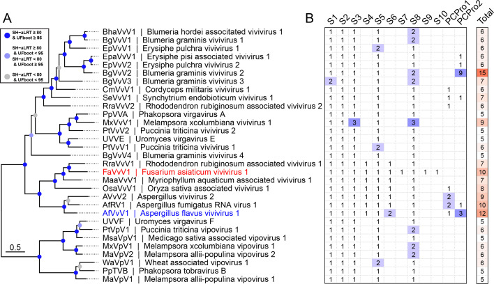
The discovery of diverse segmented RNA viruses through metatranscriptomics has enabled researchers to trace their evolutionary trajectories. However, this effort has been hindered by the limited availability of complete genome sequences and the low similarity of novel viral segments. In this study, we characterized Fusarium asiaticum vivivirus 1 (FaVvV1), a +ssRNA mycovirus with 10 monocistronic RNA segments (S1 to S10, encoding VP1 to VP10), present in the phytopathogenic fungus Fusarium asiaticum. VP1 and VP2 exhibit homology with replication proteins of martellivirals, while VP3 and VP5 share similarities with the nuclear inclusion protein a and the cylindrical inclusion helicase of potyvirids, respectively. FaVvV1 forms rod-shaped virions, with VP8 functioning as a structural protein resembling the helical capsid of potyvirids and closterovirids. To explore the conservation and evolution of viviviruses, we mined 23 public Sequence Read Archive (SRA) datasets, identifying 29 vivivirus-related viruses (vivivirids) comprising 186 viral segments. VP1 (methyltransferase and RdRP domain), VP2 (methyltransferase and superfamily 1 helicase domain), VP3 (chymotrypsin-type serine protease domain), VP5 (superfamily 2 helicase domain), and VP8 (helical capsid) were identified as conserved hallmark proteins of viviviruses. Phylogenetic and structural analyses suggest that multiple genome segmentations and gene/domain duplications were involved in the evolution of vivivirids. VP3, VP5, and VP8 might share a common ancestor with potyvirids. These findings highlight the intricate evolutionary mechanisms underlying segmented virus diversity and adaptation.

Importance: Metaviromics has greatly expanded our understanding of viral diversity, including segmented or multipartite RNA viruses with genomes composed of multiple segments. However, virome analyses often fail to detect genomic segments beyond the RdRP, likely due to their low similarity to known viruses. We characterized a group of segmented, potentially multipartite, +ssRNA viruses, with Fusarium asiaticum vivivirus 1 as a representative; most of these viruses likely infect fungi. Through structural and evolutionary analysis of the five core segments of viviviruses, our findings highlight key aspects of vivivirus evolution, including genome segmentation, gene and domain duplications, and segments with multiple evolutionary origins.

Keywords: genome segmentation; mycovirus; rod-shaped virion; segmented virus.


Conflict of interest statement

The authors declare no conflict of interest.

Figures

Fig 1
Fig 1
Genome features and virion of FaVvV1. (A) The 10 complete genomic segments of FaVvV1. Putative ORFs are presented as white arrows, and the protein domains and annotations are marked using colored rectangles. Regions with colored dashed lines indicate high nucleotide similarity among RNA segments S8, S9, and S10, with length and similarity information shown in boxes. Predicted papain-like cysteine protease cleavage sites in VP4 and VP6 are indicated by arrows. Mtr1, Methyltransferase (Mtr) domain in VP1. Mtr2, Mtr domain in VP2. RdRP, RNA-dependent RNA polymerase. SF1H, superfamily 1 helicase. SF2H, superfamily 2 helicase. CSPro, chymotrypsin-type serine protease. PCPro, papain-like cysteine protease. CP, capsid. The figure was drawn using the R package gggenes 0.5.1 (https://cran.r-project.org/web/packages/gggenes). (B) Alignment of the 5′ terminal region of FaVvV1. (C) Alignment of the 3′ terminal regions of FaVvV1. Long poly(A) tails at the 3′ termini were trimmed for alignment. Residues with >50% identity are colored. (D) Morphology of FaVvV1 virions. (E) SDS-PAGE of FaVvV1 structural proteins. (F) FaVvV1 virion length distribution. (G) Peptide distribution of the predicted capsid ORF. The first peptide match location is marked with a dotted line. (H) Structural alignment of the capsids of FaVvV1 and sweet potato mild mottle virus (SPMMV). Left, ColabFold2-predicted structure of FaVvV1 capsid, colored according to the pLDDT score. Center, coat protein of sweet potato mild mottle virus (PDB: 8ACC) in wheat color (37). Right, superposition of the two structures with TM-align.
Fig 2
Fig 2
Newly discovered viviviruses and vipoviruses. (A) Phylogenetic analysis of assembled viruses based on the Mtr1-RdRP domain. FaVvV1 described here is highlighted in red. Another recently reported vivivirus, AfVvV1, with a complete genome is in blue. The best-fit model is Q.pfam + F + I + G4 according to the Bayesian information criterion. For better clarity, nodes are color-coded based on their branch support values: nodes with SH-aLRT ≥80 and UFboot ≥95 are marked in blue, those with either SH-aLRT ≥80 and UFboot <95 or SH-aLRT <80 and UFboot ≥95 are shown in light blue, and branches where both SH-aLRT <80 and UFboot <95 are displayed in gray. (B) Genome contents and RNA segment numbers of assembled viviviruses and vipoviruses with FaVvV1 as a reference. PCPro1 and PCPro2 refer to homologous RNA segments containing a papain-like cysteine protease, excluding PCPro-containing segments S4 and S6.
Fig 3
Fig 3
Comparison of Mtr1 and Mtr2 domains. (A) Alignment of Mtr domains in S1 and S2 of vivivirus. Only the consensus and core sequences of the Mtr domain are shown here due to space limitations. Motifs involved in capping activity are highlighted in red. (B) The 3D structures of the Mtr domains of FaVvV1 are aligned to the known viral methyltransferase nsP1 of Chikungunya virus. Structural similarity of Mtr1 and Mtr2 compared to CHIKV_nsP1 is noted as TM-score (template modeling score) and RMSD (root mean square deviation), as calculated using jFATCAT-flexible (42). The structures of Mtr1 and Mtr2 are colored according to the value of predicted local distance difference test (pLDDT), and the structure of CHIKV_nsP1 (PDB: 7 X 01_A) is colored using a rainbow scheme (N-terminus in blue).
Fig 4
Fig 4
Phylogenetic analysis of RdRP (A), SF1H (B), and Mtr (C) domains encoded by S1 and S2 of vivivirids and pucciniviruses. All trees are midpoint-rooted. S1 and S2 of FaVvV1 are highlighted with a red five-pointed star. The “PCPro” subclade refers to a group of vivivirus-shared segments encoding papain-like cysteine protease. The best-fit models of RdRP, SF1H, and Mtr were Q.pfam + F + R9, Q.pfam + F + R8, and Q.pfam + F + R8, respectively, according to Bayesian information criterion. The complete tree is presented in .
Fig 5
Fig 5
CSPro of VP3 and SF2H of VP5 share homology with potyvirid protein. (A) AlphaFold2-predicted 3D structure of VP3. The structure is colored according to the pLDDT values. CSPro, chymotrypsin-type serine protease. (B) The chymotrypsin-type serine protease fold in S3. The putative catalytic triad of CSPro is displayed in stick and colored by elements. The structures in (B) and (C) are colored in a rainbow scheme with the N terminus in blue. (C) Phylogenetic analysis of the CSPro domain. The best-fit model was Q.pfam + I + R5. The support node annotations are the same as those in Fig. 4. The complete tree with all branches is shown in . (D) Phylogenetic analysis of the SF2H domain of FaVvV1. The model of substitution was Q.pfam + F + R7. The putative cellular homologs of the SF2H domain and ATP-dependent helicase HrpB were defined as the outgroup. The support node annotations are the same as those in Fig. 4. The complete tree with all branches is shown in Fig. S9. (E) Genome diagram of a potyvirus sweet potato mild mottle virus (SPMMV), FaVvV1, and AfVlV1. Dashed lines indicate the homologous domains.
Fig 6
Fig 6
Expansion of papain-like cysteine protease domains in viviviruses. (A) The 3D structure of the PCPro domain of VP4 and VP6 as predicted using AlphaFold2. The structure of PCPro in VP4 and VP6 is colored according to the pLDDT value. The putative catalytic triads of PCPro are displayed in stick and colored by elements. The structure of HC-Pro of turnip mosaic virus is colored in a rainbow scheme, with the N terminus in blue. PCPro, papain-like cysteine protease. HC-Pro, helper-component protease. (B) Phylogenetic analysis of the PCPro domain. The best-fit model was WAG + F + R4. The annotations are the same as those in Fig. 4. (C) Alignment of the PCPro domain. The catalytic dyad and cleavage sites are marked using a number sign (#) and asterisk (*) at the top, respectively. The full alignment is shown in Fig. S8.
Fig 7
Fig 7
Effects of FaVvV1 on F. asiaticum. (A) Colony morphology (front and rear of plates) of FaVvV1-related strains. Scale bar, 1 cm. (B) RT-PCR detection of FaVvV1 and FaMV1. BZ6, a wild-type strain co-infected with both viruses; BZ6VF, a derived strain of BZ6 with FaVvV1 eliminated. NTC, no template control. (C) Growth rates of strains BZ6 and BZ6VF on PDA. Bars represent mean ± SD, and the P-value was calculated using an unpaired two-tailed t-test. (D) Wheat heads of Zhengmai 9023 inoculated with conidia and mycelium plugs of BZ6 and BZ6VF. The inoculated spikelet was marked with black dots. Scale bar, 1 cm. The boxplot displays individual data points (dots), the median (middle line), the 25th and 75th percentiles (box), and the lower and upper whiskers, which extend from the 25th and 75th percentiles to the smallest and largest value within 1.5 × interquartile range (IQR). P-value was calculated using unpaired two-tailed t-test.

Similar articles

Cited by

References

    1. Hough B, Steenkamp E, Wingfield B, Read D. 2023. Fungal viruses unveiled: a comprehensive review of mycoviruses. Viruses 15:1202. doi: 10.3390/v15051202 - DOI - PMC - PubMed
    1. Khan HA, Nerva L, Bhatti MF. 2023. The good, the bad and the cryptic: the multifaceted roles of mycoviruses and their potential applications for a sustainable agriculture. Virology (Auckl) 585:259–269. doi: 10.1016/j.virol.2023.07.004 - DOI - PubMed
    1. Sato Y, Suzuki N. 2023. Continued mycovirus discovery expanding our understanding of virus lifestyles, symptom expression, and host defense. Curr Opin Microbiol 75:102337. doi: 10.1016/j.mib.2023.102337 - DOI - PubMed
    1. Milgroom MG, Cortesi P. 2004. Biological control of chestnut blight with hypovirulence: a critical analysis. Annu Rev Phytopathol 42:311–338. doi: 10.1146/annurev.phyto.42.040803.140325 - DOI - PubMed
    1. Wagemans J, Holtappels D, Vainio E, Rabiey M, Marzachì C, Herrero S, Ravanbakhsh M, Tebbe CC, Ogliastro M, Ayllón MA, Turina M. 2022. Going viral: virus-based biological control agents for plant protection. Annu Rev Phytopathol 60:21–42. doi: 10.1146/annurev-phyto-021621-114208 - DOI - PubMed

LinkOut - more resources