. 2025 Jul 22;99(7):e0058125.
doi: 10.1128/jvi.00581-25. Epub 2025 Jun 4.

Interferon-stimulated gene MCL1 inhibits foot-and-mouth disease virus replication by modulating mitochondrial dynamics and autophagy

Affiliations

Interferon-stimulated gene MCL1 inhibits foot-and-mouth disease virus replication by modulating mitochondrial dynamics and autophagy

Aishwarya Mogulothu et al. J Virol. .

Abstract

Interferons (IFNs) and the IFN-stimulated genes (ISGs) that they induce are effective in reducing the replication of foot and mouth disease virus (FMDV). The use of a high-throughput ISG screen identified the ISG myeloid cell leukemia 1 (MCL1) as an ISG with an antiviral effect against an FMDV replicon system. In this study, we demonstrated that overexpression of MCL1 inhibits FMDV replication by reducing approximately 4 logs of virus titers in porcine cells. We then explored the regulatory pathways associated with MCL1 to determine the specific antiviral mechanisms against FMDV. Our findings indicated that the antiviral mechanism does not involve apoptosis regulation or alterations in cell cycle phase heterogeneity. Analysis of mitochondrial function, through measurement of mitochondrial oxygen consumption rate, demonstrated that overexpression of MCL1 results in increased mitochondrial respiration and ATP production, whereas FMDV infection reduces both processes. Moreover, MCL1 overexpression resulted in elongated mitochondrial morphology, contrasting with the fragmented and punctate morphology observed during FMDV infection. Importantly, these changes in mitochondrial dynamics were independent of MCL1's regulation of mitochondrial calcium flux. We also found that MCL1 overexpression suppresses autophagy, which is known to be necessary for FMDV replication. Our data indicate that MCL1 is a potent antiviral ISG against FMDV and highlight the importance of mitochondrial dynamics and autophagy in FMDV replication.IMPORTANCEIn this study, we have successfully used a high-throughput ISG screening approach to measure the inhibition of FMDV replication using an RNA replicon system for the first time. This screen led to the identification of the potent antiviral effects of a relatively lesser-known ISG called MCL1. Our findings reveal that MCL1 exerts its antiviral functions through the regulation of mitochondrial dynamics and autophagy. Although mitochondrial dynamics are involved in apoptosis, metabolism, redox homeostasis, stress responses, and antiviral signaling, this pathway has not been thoroughly explored in the context of FMDV infection. Further investigation into mitochondrial dynamics may facilitate the development of improved biotherapeutics for FMDV. Additionally, our studies highlight the significance of autophagy, a pathway that is needed by FMDV for replication. Ultimately, a deep understanding of all mechanisms exploited by FMDV may allow for the rational design of novel therapeutics and vaccines to control FMD.

Keywords: antiviral agents; autophagy; foot-and-mouth disease virus; interferon-stimulated genes; interferons; mitochondrial metabolism.


Conflict of interest statement

The authors declare no conflict of interest.

Figures

Fig 1
Fig 1
FMDV replication in MCL1-overexpressing porcine cells. (A) MGPK-αvβ6 cells were transduced with SCRPSY lentivirus either expressing GFP, huMCL1, or porMCL1. They were then infected with FMDV A12 WT at an MOI of 5 and followed by a 6 h incubation period (n = 3). Virus titers were determined by plaque assay on BHK-21 cells. (B) Western blot was conducted on lysates collected at 6 h post-infection and probed with antibodies against MCL1, FMDV VP1, and actin. Statistical analysis was performed using one-way ANOVA with the Tukey post hoc test.
Fig 2
Fig 2
FMDV replication in porcine cells treated with Q-VD-OPH. (A) MGPK-αvβ6 cells were treated with DMSO, 1 µM of staurosporine (STS), 20 µM of Q-VD-OPH (QVD) and STS, FMDV A12 WT MOI: 5, or QVD and A12 WT. Lysates were collected at indicated time points and run on a western blot to be probed with antibodies against PARP, actin, or FMDV VP1. (B) MGPK-αvβ6 cells were treated with either DMSO or 20 µM of QVD, followed by infection with FMDV A12 WT at an MOI: 5 (n = 3). Virus titers were determined by plaque assay on BHK-21 cells. Statistical analysis was performed using 2-way ANOVA with the Tukey post hoc test.
Fig 3
Fig 3
Cell cycle analysis of MCL1 overexpressing porcine cells. MGPK-αvβ6 cells overexpressing either GFP or porMCL1 were collected, fixed, and then stained with DAPI before being analyzed by flow cytometry. (A) Representation of the cell counts in each phase of the cell cycle (G0/G1, S, G2/M) based on DAPI fluorescence for both overexpressing cell lines. (B) Percentage (%) distribution of each cell cycle phase in MGPK-GFP and MGPK-porMCL1 cells. Statistical analysis was performed using Student’s t-test.
Fig 4
Fig 4
Mito stress test on porcine cells overexpressing MCL1. (A) Mito stress test was conducted with a Seahorse bioanalyzer on MGPK-GFP and MGPK-porMCL1 cells that were mock-infected or infected with FMDV A12 WT MOI: 5. Subsequent addition of oligomycin, FCCP, and Rotenone/antimycin A allowed for calculation of different types of respiration occurring in mitochondria. (B) Basal respiration-related OCR from mito stress test in (A). Basal respiration is oxygen consumption under basal conditions to meet the ATP demand of the cells, and also due to proton leak. (C) ATP production OCR from mito stress test in (A). ATP production is oxygen consumption required to create ATP and is measured after the addition of oligomycin. (D) Coupling efficiency from mito stress test from (A). Coupling efficiency refers to how efficiently mitochondria are at consuming oxygen, coupled to ATP production versus proton leak. It is calculated by dividing ATP-linked OCR by basal respiration-linked OCR. Statistical analysis was performed using one-way ANOVA with the Tukey post hoc test.
Fig 5
Fig 5
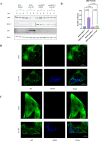
Mitochondrial morphology of porcine cells overexpressing MCL1. MGPK-EMPTY (A) and MGPK-porMCL1 (B) cells were either mock-infected or infected with FMDV A12 WT MOI: 5 and were fixed at 4 h post-infection. The proteins were visualized by indirect immunofluorescence. HSP60 was detected with a rabbit monoclonal antibody and then with Alexa Fluor 488-conjugated secondary antibody. RFP was an indicator for lentivirus-transduced cells (not shown). Double-stranded RNA (dsRNA) was detected with a mouse monoclonal antibody and then with Alexa Fluor 405-conjugated secondary antibody. The scale bar is representative of 10 µm. (C) Analysis of mitochondrial networks was done with Fiji (n = 5). The average number of networks graph is representative of all mitochondrial networks that contain one or more branches. The network size is indicative of an average number of branches per mitochondrial network. Statistical analysis was performed using one-way ANOVA with the Tukey post hoc test.
Fig 6
Fig 6
Mitochondrial calcium levels of porcine cells overexpressing MCL1. MGPK-αvβ6 cells were transfected with pcDNA3.1(+) or pcDNA3.1(+)-porMCL1 plasmids and then were mock-infected or infected with A12 WT MOI: 5 for 6 h. The cells were then stained with rhod 2-AM, viability dye, fixed, permeabilized, and finally stained with a monoclonal antibody for MCL1 that was conjugated to APC before running flow cytometry. (A) The figure represents the gating strategy for cells transfected with pcDNA 3.1(+), and (B) is for pcDNA 3.1(+)-porMCL1. The gating strategy employed segregation of cells overexpressing MCL1 (MCL1(+)) and cells endogenously expressing MCL1 (MCL1(-)). Rhod 2-AM was measured on the PE channel. (C) Representations of the median PE-H for both MCL1(+) and (D) MCL1(-) populations in the infected and uninfected samples. Statistical analysis was done using one-way ANOVA with the Tukey post hoc test.
Fig 7
Fig 7
Analysis of autophagic flux with p62 puncta formation in porcine cells overexpressing MCL1. (A) Western blot analysis of lysates from MGPK-GFP and MGPK-porMCL1 cells subjected to a mock infection or infected with FMDV A12 WT at an MOI of 5 over a time course. The western blot was probed with antibodies against autophagic marker p62, MCL1, FMDV VP1, and actin as a loading control. (B) Indirect immunofluorescence imaging of p62 and dsRNA signals in MGPK-EMPTY cells compared with cells that were infected with FMDV A12 WT MOI: 5 for 6 h. (C) Indirect immunofluorescence imaging of MGPK-porMCL1 cells that were either mock-infected or infected for 6 h with FMDV A12 WT MOI: 5. The p62 protein was visualized using a polyclonal rabbit antibody and a secondary Alexa Fluor 488 antibody. The presence of dsRNA was visualized using a monoclonal antibody and a secondary Alexa Fluor 405 antibody. RFP was an indicator for lentivirus-transduced cells (not shown). The scale bar is representative for 10 µm. (D) Analysis of the number of p62 puncta was done with Fiji, and the graph represents the average number of puncta for each sample type (n = 5). Statistical analysis was performed using one-way ANOVA with the Tukey post hoc test.
Fig 8
Fig 8
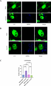
Analysis of autophagic flux with LC3 puncta formation in porcine cells overexpressing MCL1. MGPK-EMPTY (A) and MPGK-porMCL1 (B) cells were either mock-infected or infected with FMDV A12 WT MOI: 5 and were fixed 6 hours post-infection. The proteins were visualized using indirect immunofluorescence. LC3 was detected using a rabbit monoclonal antibody and a secondary antibody conjugated with Alexa Fluor 488. The presence of dsRNA was detected using a mouse monoclonal antibody and a secondary antibody conjugated with Alexa Fluor 405. RFP was an indicator for lentivirus-transduced cells (not shown). The scale bar is representative of 10 µm. (C) Analysis of the number of LC3 puncta was done with Fiji, and the graph represents the average number of puncta for each sample type (n = 5). Statistical analysis was performed using one-way ANOVA with the Tukey post hoc test.
Fig 9
Fig 9
Expression of ISG15 mRNA in response to stimulation with poly(I:C) or A12 WT infection. Total RNA was isolated in MGPK-GFP and MGPK-porMCL1 cells that were either treated with poly(I:C) (A) or infected with A12 WT MOI: 5 (B) to analyze the mRNA fold expression of ISG15 at 6 h post-infection by real-time RT-PCR. Porcine HRP-T was used as an internal control, and all the results were expressed as a relative fold increase in gene expression with respect to mock-treated cells (n = 3). Statistical analysis was performed using Student’s t-test.
Fig 10
Fig 10
Effect of MCL1 and FMDV on mitochondrial dynamics and autophagy. This model illustrates the contrasting effects of MCL1 overexpression and FMDV infection on mitochondrial dynamics and autophagy in porcine cells. MCL1 overexpression in porcine cells leads to increased mitochondrial fusion, leading to increased oxidative phosphorylation, resulting in increased ATP production and highly networked mitochondria. On the other hand, FMDV leads to mitochondrial fragmentation, leading to decreased oxidative phosphorylation and associated ATP production, as well as the formation of punctate mitochondria. Additionally, FMDV triggers autophagy and exploits the autophagic machinery to facilitate its viral replication. MCL1, by inhibiting autophagy, reduces the availability of this machinery for viral replication, ultimately leading to decreased FMDV replication. Created in BioRender.com

Similar articles

References

    1. Grubman MJ, Baxt B. 2004. Foot-and-mouth disease. Clin Microbiol Rev 17:465–493. doi: 10.1128/CMR.17.2.465-493.2004 - DOI - PMC - PubMed
    1. World Organisation for Animal Health . 2022. Foot and mouth disease. WOAH - World Organisation for Animal Health. Available from: https://www.woah.org/en/disease/foot-and-mouth-disease
    1. World Organisation for Animal Health . 2022. Terrestrial code online access. WOAH - World Organisation for Animal Health. Available from: https://www.woah.org/en/what-we-do/standards/codes-and-manuals/
    1. Medina GN, de Los Santos T, Diaz-San Segundo F. 2020. Use of IFN-based biotherapeutics to harness the host against foot-and-mouth disease. Front Vet Sci 7:465. doi: 10.3389/fvets.2020.00465 - DOI - PMC - PubMed
    1. Schoggins JW, Wilson SJ, Panis M, Murphy MY, Jones CT, Bieniasz P, Rice CM. 2011. A diverse range of gene products are effectors of the type I interferon antiviral response. Nature 472:481–485. doi: 10.1038/nature09907 - DOI - PMC - PubMed

MeSH terms

Substances

LinkOut - more resources