. 2025 Jun 4:10.1007/s10753-025-02320-6.
doi: 10.1007/s10753-025-02320-6. Online ahead of print.

Activation of Human FPR2 with AT-RvD1 Resolves Acute Sialadenitis in Vivo

Affiliations

Activation of Human FPR2 with AT-RvD1 Resolves Acute Sialadenitis in Vivo

Kihoon Nam et al. Inflammation. .

Abstract

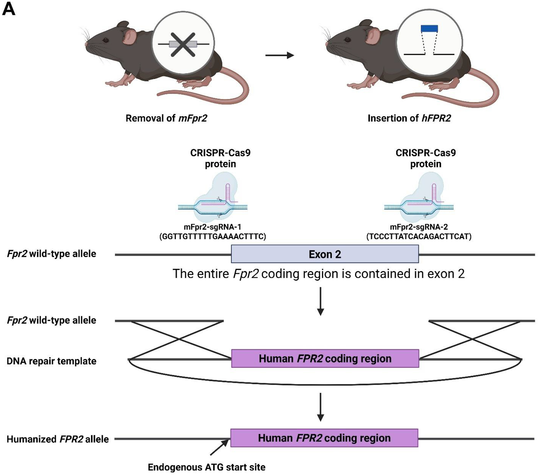
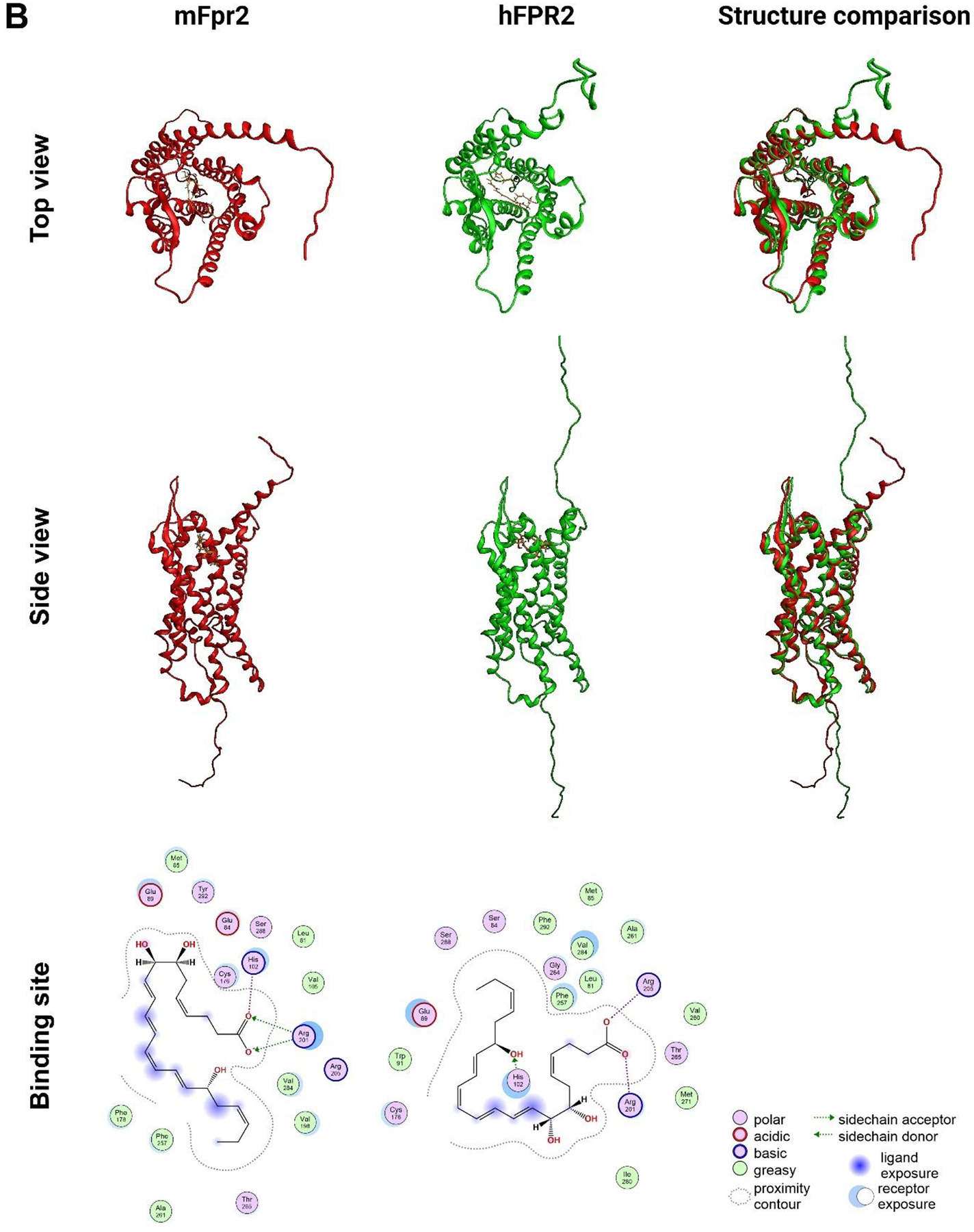
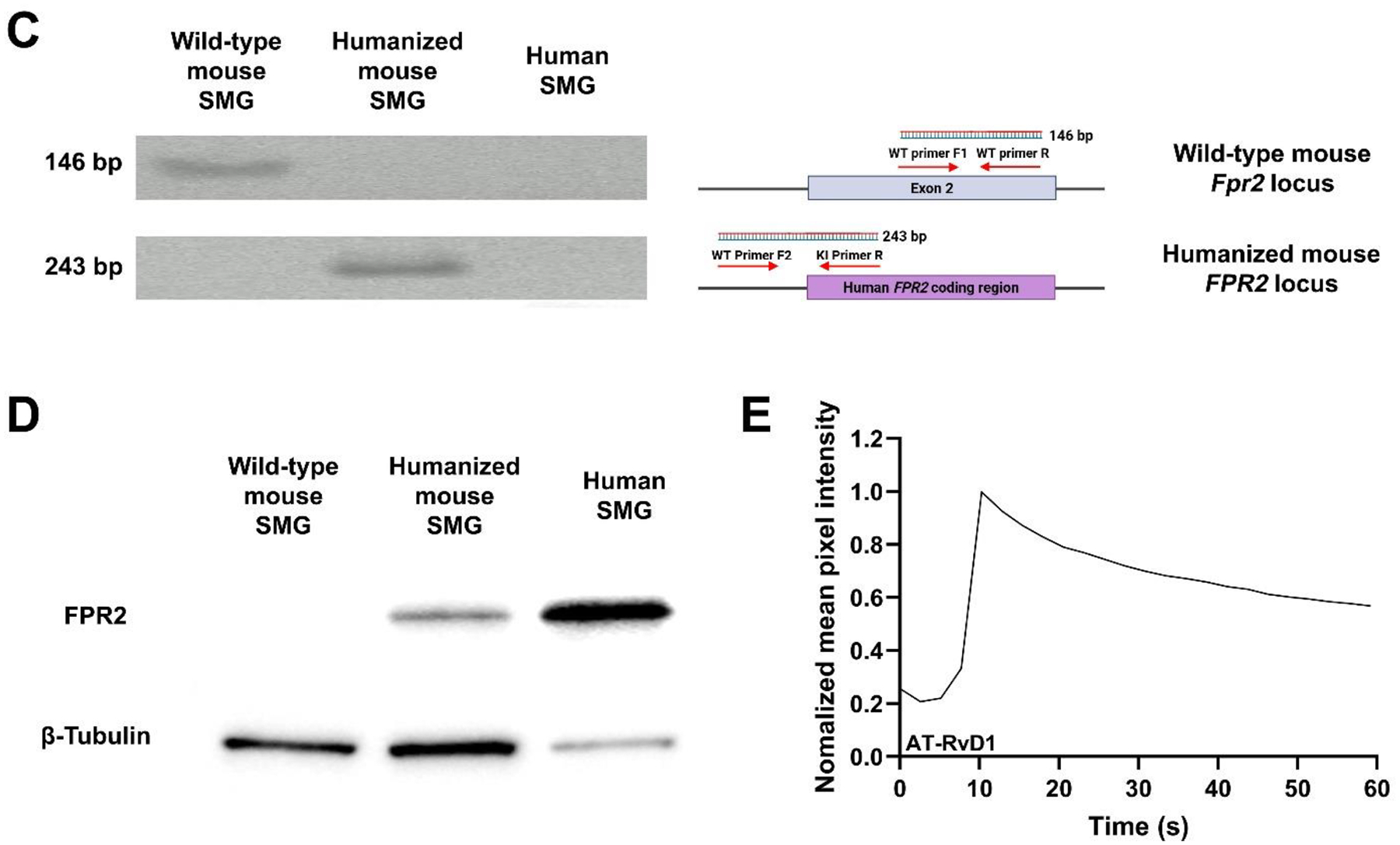
Previous studies demonstrated that activation of the mouse G protein-coupled formyl peptide receptor 2 (mFpr2) with aspirin-triggered resolvin D1 (AT-RvD1) blocks pro-inflammatory cytokine signaling while promoting salivary gland (SG) epithelial integrity both in vitro and in vivo. In addition, mice lacking Fpr2 display alterations of SG innate and adaptive immunity. Taken together, these results indicate that Fpr2 activation with AT-RvD1 restores saliva secretion and regulates SG immunity in mice. To demonstrate the value of AT-RvD1 for use in human SG, however, we need to extend the findings above in the direction of clinical use. To this end, the current study investigated whether treatment with AT-RvD1 reduces SG inflammation and restores saliva secretion in an acute sialadenitis mouse model expressing the human formyl peptide receptor 2 (hFPR2) protein. Results indicate that mice carrying the hFPR2 and treated with lipopolysaccharide (LPS) display acute sialadenitis-like features as shown by increased levels of proliferating inflammatory cells, loss of epithelial integrity and reduced saliva secretion. In contrast, when these mice are treated with AT-RvD1, the sialadenitis-like features are drastically reduced as evidenced by a significant decrease in proliferating inflammatory cells as well as restoration of saliva secretion to levels comparable to phosphate buffered saline (PBS)-treated healthy controls. Finally, changes observed in mice carrying the hFPR2 and treated with LPS and AT-RvD1 were comparable to those observed in wild-type mice carrying the mFpr2. Together, these results demonstrate that activation of hFPR2 with AT-RvD1 resolves acute sialadenitis in vivo.

Keywords: Immunity; Inflammation; Lipids; Oral pathology; Receptors; Salivary physiology.


Conflict of interest statement

Declarations. Competing interest: The authors declare no competing interests. Ethical Approval: All animal handling, anesthesia, and treatments were conducted in compliance with Animal Research: Reporting of In Vivo Experiments guidelines and received prior approval from the University of Missouri-Columbia Animal Care and Use Committee (ACUC), approval number: 42063. Moreover, all human specimen analyses were conducted under the guidelines and with the approval of the University of Missouri-Columbia Institutional Review Board (IRB), approval number: 2024461, with informed consent obtained for each patient collected from their medical record.

Figures

Figure 1.
Figure 1.. Generation and characterization of humanized FPR2 knock-in mice.
A DNA repair template was used to replace the mouse Fpr2 coding region with the human FPR2 sequence, thereby integrating it at the endogenous ATG start site (A). Structural predictions of mouse Fpr2 (red) and human FPR2 (green) were generated using ColabFold v1.5.5 and visualized with the PyMOL Molecular Graphics System (version 3.0). Ligand (AT-RvD1, orange molecule) interactions were modeled using the Molecular Operating Environment (MOE) software. The resulting models revealed overall structural similarity between the two receptors but likewise revealed differences in ligand binding site composition (B). Successful replacement of the mouse Fpr2 gene with the human FPR2 coding sequence was confirmed by PCR performed on cDNA synthesized from total RNA extracted from wild-type mice, humanized mice and human SMG tissue with the wild-type transcript (146 bp) amplified using a forward primer (WT primer F1) and a reverse primer (WT primer R), while the humanized transcript (243 bp) was detected using a knock-in-specific forward primer (WT primer F2) and a reverse primer located within the human FPR2 sequence (KI primer R) (C). Expression of the human FPR2 protein was confirmed by Western blot analysis using an antibody that selectively binds human FPR2. The protein was detected in humanized mouse and human SMG but not in wild-type mouse SMG, with β-tubulin used as a loading control in each of these cases (D). Functional expression of hFPR2 was validated by calcium imaging. SMG cells were loaded with Fura-2 AM and stimulated with AT-RvD1 (100 ng/mL). Intracellular calcium levels ([Ca2+]i) increased rapidly upon stimulation, indicating intact receptor function. Data shown are representative of n = 12 (E).
Figure 2.
Figure 2.. AT-RvD1 restores morphology in SMG from hFPR2 mice treated with LPS.
Rehydrated SMG tissue sections from humanized FPR2 mice treated with LPS (A, E), AT-RvD1 + LPS (B, F) and PBS (C, G) were stained with hematoxylin-eosin and images captured using a Leica DMI6000B. Results show that SMG treated with LPS display a loss of epithelial integrity together with areas of acinar shrinkage (white arrows). In contrast, SMG treated with AT-RvD1 + LPS showed restoration of tissue architecture as evidenced by a reduction of acinar shrinkage compared to those with LPS alone. To quantify these changes, tissue sections were stained with goat anti-Muc10 and mouse anti-E-Cadherin and counterstained with DAPI (blue). Next, acinar cell size for each treatment was calculated using ImageJ (D) and expressed as mean ± SD of results from a total of 4 mice per group in quadruples. Statistical significance was assessed by one-way ANOVA (where *p ≤ 0.05 is considered statistically significant) followed by Dunnett’s post-hoc test for multiple comparisons (H). Scale bars represent 50 μm.
Figure 3.
Figure 3.. AT-RvD1 reduces proliferating inflammatory cells in SMG from hFPR2 mice treated with LPS.
SMG tissue sections from hFPR2 mice treated with LPS (A), AT-RvD1 + LPS (B) and PBS (C) were analyzed using confocal microscopy with rabbit anti-Ki67 (green) and rat anti-CD45 (red) antibodies, and counterstained with DAPI (blue). Purple arrows indicate Ki67-only positive cells, while yellow arrows indicate CD45-only positive cells and light white arrows indicate Ki67 and CD45 double-positive cells, respectively. Our results show that SMG treated with LPS displayed an increased number of proliferating cells (D), CD45+ cells (E), and proliferating CD45+ cells (F). In contrast, SMG treated with AT-RvD1 + LPS showed a significant reduction in proliferating cells (D), CD45+ cells (E), and proliferating CD45+ cells (F). Samples were examined using a Leica STELLARIS 5 confocal microscope equipped with an artificial intelligence-guided image analysis software (AIVIA). Representative image from a total of 4 mice per group in quadruples. Statistical significance was assessed by one-way ANOVA (where *p ≤ 0.05 is considered statistically significant) followed by Dunnett’s post-hoc test for multiple comparisons (D-F). Scale bars represent 50 μm.
Figure 4.
Figure 4.. AT-RvD1 restores epithelial polarization in SMG from hFPR2 mice treated with LPS.
SMG tissue sections from hFPR2 mice treated with LPS (A, E; red line), AT-RvD1 + LPS (B, E; blue line) and PBS (C, E; black line) were analyzed using confocal microscopy with rabbit anti-ZO-1 (green) and mouse anti-E-Cadherin (red) antibodies, and counterstained with DAPI (blue). Our results show that SMG that were treated with LPS displayed a significant reduction in ZO-1 expression when compared to PBS treated healthy controls (F), note decreased intensity in some areas (A; white arrows). In contrast, SMG treated with AT-RvD1 + LPS showed a significant increase in ZO-1 expression (F). Samples were examined using a Leica STELLARIS 5 confocal microscope and analyzed using ImageJ as described in Methods (D). Representative image from a total of 4 mice per group in quintuples. Statistical significance was assessed by one-way ANOVA (where *p ≤ 0.05 is considered statistically significant) followed by Dunnett’s post-hoc test for multiple comparisons (F). Scale bars represent 50 μm.
Figure 5.
Figure 5.. AT-RvD1 restores Aquaporin-5 expression in SMG and saliva secretion from hFPR2 mice treated with LPS.
SMG tissue sections from hFPR2 mice treated with LPS (A, D, G; red line), AT-RvD1 + LPS (B, E, G; blue line) and PBS (C, F, G; black line) were analyzed using confocal microscopy with rabbit anti-Aquaporin-5 (AQP5, green) and mouse anti-Ki67 (A-C; red) or CD45 (D-F; red) antibodies, and counterstained with DAPI (blue). Our results show that SMG treated with LPS displayed a local reduction in apical AQP5 expression as compared to PBS treated healthy controls (white dotted circle areas) and a significant translocation of this protein toward the basolateral area in zones with CD45+ cells (white arrows) (H). In contrast, SMG treated with AT-RvD1 + LPS showed a significant increase in apical AQP5 signal. Samples were examined using a Leica STELLARIS 5 confocal microscope and analyzed using ImageJ as described in Methods. Results are expressed as mean ± SD of results from a total of 4 mice per group in quintuples. Statistical significance was assessed by one-way ANOVA (where *p ≤ 0.05 is considered statistically significant) followed by Dunnett’s post-hoc test for multiple comparisons (H). After two days from the treatment, mice were anesthetized, and saliva secretion was induced with pilocarpine using a total of 7 mice per group. Statistical significance was assessed by one-way ANOVA (where *p ≤ 0.05 is considered statistically significant) followed by Dunnett’s post-hoc test for multiple comparisons. The saliva flow rate from humanized FPR2 mice treated with LPS was significantly reduced. In contrast, mice treated with AT-RvD1 + LPS showed a significant increase in saliva flow rate (I). Scale bars represent 50 μm.

References

    1. McQuone SJ, Acute viral and bacterial infections of the salivary glands. Otolaryngol Clin North Am, 1999. 32(5): p. 793–811. - PubMed
    1. Moore J, et al. , Approach to sialadenitis. Can Fam Physician, 2023. 69(8): p. 531–536. - PMC - PubMed
    1. Ramazani F, et al. , Development of a patient reported outcome instrument for chronic sialadenitis. J Otolaryngol Head Neck Surg, 2022. 51(1): p. 4. - PMC - PubMed
    1. Nelson JW, et al. , ALX/FPR2 receptor for RvD1 is expressed and functional in salivary glands. Am J Physiol Cell Physiol, 2014. 306(2): p. C178–85. - PMC - PubMed
    1. Odusanwo O, et al. , Resolvin D1 prevents TNF-alpha-mediated disruption of salivary epithelial formation. Am J Physiol Cell Physiol, 2012. 302(9): p. C1331–45. - PMC - PubMed

LinkOut - more resources