Macrophage mannose receptor CD206-targeted PET imaging in experimental acute myocardial infarction
- PMID: 40465093
- PMCID: PMC12137864
- DOI: 10.1186/s13550-025-01254-2
Macrophage mannose receptor CD206-targeted PET imaging in experimental acute myocardial infarction
Abstract
Background: The macrophage mannose receptor (CD206) is expressed predominantly on the surface of M2-type macrophages, which play a role in resolution of inflammation after myocardial injury. The purpose of this study was to evaluate the utility of CD206-targeted PET tracer Al[18F]F-NOTA-D10CM, a fluorinated mannosylated dextran derivative, for imaging immune responses after experimental acute myocardial infarction (MI).
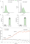
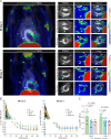
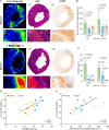
Results: Flow cytometry revealed selective binding of Alexa-488-NOTA-D10CM to human M2-polarized macrophages derived from blood monocytes compared to M1 macrophages. The binding affinity of Al[18F]F-NOTA-DCM for CD206-positive Chinese hamster ovary cells was 1.83 ± 0.68 nM. In vivo PET and ex vivo autoradiography experiments in Sprague-Dawley rats studied at 3 and 7 days after permanent ligation of the left coronary artery or a sham-operation, showed significantly higher uptake of Al[18F]F-NOTA-DCM in the MI area than in remote areas, or the myocardium of sham-operated rats. However, there was no difference in uptake in the MI area between day 3 and day 7. Uptake of Al[18F]F-NOTA-DCM in the MI area correlated positively with the area-% of CD206-positive staining of the left ventricular myocardium (r = 0.481, P = 0.006). In vitro competition studies on tissue cryosections using a molar excess of unlabeled D10CM revealed a reduction of approximately 85%, confirming specific tracer binding.
Conclusion: Al[18F]F-NOTA-D10CM PET detects overexpression of CD206 after ischemic myocardial injury, and may be a suitable biomarker for detecting M2-type macrophages associated with the inflammatory process post-MI.
Keywords: CD206; Inflammation; Macrophage mannose receptor; Myocardial infarction; PET.
© 2025. The Author(s).
Conflict of interest statement
Declarations. Ethics approval: All animal experiments were approved by the national Project Authorization Board in Finland (license number ESAVI/43134/2019) and carried out in compliance with the EU Directive 2010/EU/63 on the protection of animals used for scientific purposes. Consent for publication: Not applicable. Competing interests: Dr. Saraste received consultancy fees from AstraZeneca and Pfizer, and speaker fees from Abbott, AstraZeneca, Janssen, Novartis, and Pfizer (not related to the current study). Dr. Knuuti received consultancy fees from GE Healthcare and Synektik, and speaker fees from Bayer, Lundbeck, Boehringer-Ingelheim, Pfizer, and Siemens (outside of the submitted work). The remaining authors have no conflicts of interest to disclose.
Figures


Similar articles
-
Macrophage mannose receptor CD206 targeting of fluoride-18 labeled mannosylated dextran: A validation study in mice.Eur J Nucl Med Mol Imaging. 2024 Jul;51(8):2216-2228. doi: 10.1007/s00259-024-06686-x. Epub 2024 Mar 27. Eur J Nucl Med Mol Imaging. 2024. PMID: 38532026 Free PMC article.
-
In vivo Visualization of M2 Macrophages in the Myocardium After Myocardial Infarction (MI) Using 68 Ga-NOTA-Anti-MMR Nb: Targeting Mannose Receptor (MR, CD206) on M2 Macrophages.Front Cardiovasc Med. 2022 Apr 25;9:889963. doi: 10.3389/fcvm.2022.889963. eCollection 2022. Front Cardiovasc Med. 2022. PMID: 35548425 Free PMC article.
-
Aluminum Fluoride-18 Labeled Mannosylated Dextran: Radiosynthesis and Initial Preclinical Positron Emission Tomography Studies.Mol Imaging Biol. 2023 Dec;25(6):1094-1103. doi: 10.1007/s11307-023-01816-7. Epub 2023 Apr 4. Mol Imaging Biol. 2023. PMID: 37016195 Free PMC article.
-
PET imaging of angiogenesis after myocardial infarction/reperfusion using a one-step labeled integrin-targeted tracer 18F-AlF-NOTA-PRGD2.Eur J Nucl Med Mol Imaging. 2012 Apr;39(4):683-92. doi: 10.1007/s00259-011-2052-1. Epub 2012 Jan 25. Eur J Nucl Med Mol Imaging. 2012. PMID: 22274731 Free PMC article.
-
PET of glucagonlike peptide receptor upregulation after myocardial ischemia or reperfusion injury.J Nucl Med. 2012 Dec;53(12):1960-8. doi: 10.2967/jnumed.112.109413. Epub 2012 Nov 8. J Nucl Med. 2012. PMID: 23139087
References
-
- Thackeray JT, Lavine K, Liu Y. Imaging inflammation past, present, and future: focus on cardioimmunology. J Nucl Med. 2023;64:39S-S48. - PubMed
-
- Park JB, Suh M, Park JY, Park JK, Kim YI, Kim H, et al. Assessment of inflammation in pulmonary artery hypertension by 68Ga-mannosylated human serum albumin. Am J Respir Crit Care Med. 2020;201:95–106. - PubMed
Grants and funding
LinkOut - more resources
Full Text Sources

