. 2025 Jun 4;20(6):e0325480.
doi: 10.1371/journal.pone.0325480. eCollection 2025.

Radioprotective effects of asiaticoside and asiatic acid in neural stem cells derived from human stem cells from apical papilla through increasing dose-reduction factor and their lowering effects on SH-SY5Y cell viability

Affiliations

Radioprotective effects of asiaticoside and asiatic acid in neural stem cells derived from human stem cells from apical papilla through increasing dose-reduction factor and their lowering effects on SH-SY5Y cell viability

Taweesak Tangrodchanapong et al. PLoS One. .

Abstract

Objective: To investigate the effects of asiaticoside (AS) and asiatic acid (AA) against radiotherapy on neural stem cells induced from human stem cells from apical papilla (NSCs-hSCAPs) through dose-reduction factor (DRF) evaluation and their radiosensitization on human neuroblastoma SH-SY5Y cells.

Methods: NSCs-hSCAPs were treated with AS or AA (0-500 μM) and radiation (0-8 Gy). Isolated hSCAPs were verified mesenchymal stem cells (MSCs) properties according to standard protocol. Subsequently, NSCs-hSCAPs were characterized by Cresyl violet staining and immunocytochemistry. A culture plate containing the cells was embedded into the solid water and bolus phantom. After CT simulation and treatment planning, dose uniformity to the plate was evaluated. X-ray, AS, and AA toxicity were investigated using cell viability (MTT) assay. Finally, DRF50 was calculated from dose-response curves at 50% cell viability for both cell lines.

Results: hSCAPs presented MSCs markers. NSCs-hSCAPs were successfully generated due to the Nissl substance, Nestin, and SOX2 positively stained. Dose homogeneity was represented as isodose at 100% covered the cells in the phantom, suggesting that they were received according to prescribed doses. MTT results revealed that AA was more toxic than AS in both cells. X-ray reduced significantly in a number of tested cells and more radiosensitivity was observed in SH-SY5Y. However, the reduction affected by 4 Gy was diminished after AA or AS at 2 μM applied to NSCs-hSCAPs. Moreover, a significant increase of DRF50 was found at 2 μM of AA (6.72 ± 2.35) and AS (3.84 ± 1.41) in NSCs-hSCAPs whereas it did not show in SH-SY5Y. Interestingly, 20 μM AA could reduce SH-SY5Y cell viability (mean of the cell viability (%) was 25.22 ± 1.53 compared to 30.22 ± 1.46 in the control group), showing a very large in terms of its effect size (Cohen's d value = 1.37).

Conclusion: AA and AS had a specific radioprotective effect on NSCs-hSCAPs without affecting SH-SY5Y. However, AA might be a better therapeutic agent due to expressing a lethal effect on the irradiated cancer cells.


Conflict of interest statement

The authors have declared that no competing interests exist.

Figures
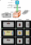
Fig 1
Fig 1. Irradiation set-up of solid water and bolus phantom and treatment planning on CT images.
(A) The diagram shows the configuration and irradiation set-up of the phantom. (B – D) Isodose coverages of PTV (96-well plate) on phantom’s CT images by using treatment planning system on axial, coronal, and sagittal plane.
Fig 2
Fig 2. Characterization of hSCAPs.
(A) The human third molar teeth presented the apical papilla tissue (white arrows) and (A’) the dissected apical papilla tissue (white arrow). (B) The isolated cells from human apical papilla tissue presented adherent ability and fibroblast-like shape (black arrows). (C – D) The ectomesenchymal origin was revealed by β-III tubulin and Nestin immunofluorescent staining. (E) Colony-forming unit (black arrows) represented their self-renewal ability. (F – H) Multi-linage differentiation abilities were demonstrated by adipogenesis (black arrows indicate lipid droplets), osteogenesis (white arrows indicate extracellular calcium nodules), and neurogenesis (black arrows indicate Nissl bodies), respectively. (I) The cell surface antigen molecules profiling of MSCs was investigated by flow cytometry. Scale bars: A, A’, E = 5 mm, B – D = 100 µm, and F – H = 50 µm.
Fig 3
Fig 3. Characterization of NSCs-hSCAPs.
The dynamic change of cell morphology of NSCs-hSCAPs. (A) The hSCAPs presented the adherent flattened-shaped cells. (B – D) Under the neural-inducing environment, cells were suspended and aggregated into the 3D-neurosphere, which consisted of intraspheroidal cells. (E) The Cresyl violet staining revealed that these intraspheroidal cells demonstrated the dark purple of the Nissl substance. (F – L) Immunofluorescence profiling of NSCs-hSCAPs under 3D neurosphere induction. (G – H) The positive expression of the early neuronal stage, Nestin and SOX2 was observed, respectively. Scale bars: A – E = 50 µm, and F – L = 100 µm.
Fig 4
Fig 4. Effect of AS, AA, and X-ray radiation on cell viability (%).
(A) The chemical structures of AS and AA. (B – D) Cell viability (%) of NSCs-hSCAPs treated with AS (0-500 µM) at 24 hours, AA (0-500 µM) at 24 hours, and X-ray (0-8 Gy) at 24 and 48 hours, respectively. (E – G) Cell viability (%) of SH-SY5Y treated with AS (0–500 µM) at 24 hours, AA (0–500 µM) at 24 hours, and X-ray (0–8 Gy) at 24 and 48 hours, respectively. Results are represented as mean cell viability (%) ± SEM from three independent experiments. The number of samples is n = 9 for each condition. Error bars represent SEM. **** = p < 0.0001 compared to the control group, ## = p < 0.01 compared to the vehicle, #### = p < 0.0001 compared to the vehicle (Fig 4B, 4C, 4E, and 4F). * = p < 0.05, **** = p < 0.0001 compared to the control group at 24 hours, ### = p < 0.001, #### = p < 0.0001 compared to the control group at 48 hours (Fig 4D and 4G).
Fig 5
Fig 5. Cell viability (%) of irradiated NSCs-hSCAPs and SH-SY5Y in AS and AA treatment.
Effect of AS and AA on X-ray radiation (6 MV)-induced reduction of cell viability (%) in NSCs-hSCAPs and SH-SY5Y cells. Cell viability (%) of the irradiated NSCs-hSCAPs (A – D) or SH-SY5Y cells (E – H) with 2 - 8 Gy in the presence of AS at non-toxic concentrations (0 - 20 µM). Cell viability (%) of the irradiated NSCs-hSCAPs (I – L) or SH-SY5Y cells (M – P) with 2 - 8 Gy in the presence of AA at non-toxic concentrations (0 - 20 µM). The cell viability (%) is represented as mean ± SEM from three independent experiments. The number of samples is n = 9 for each condition. * p < 0.05 and ** p < 0.01 vs. untreated control NSCs-hSCAPs or SH-SY5Y cells.
Fig 6
Fig 6. Dose-response curves and DRF50 of NSCs-hSCAPs and SH-SY5Y in the presence of AS and AA.
Dose-response curve of NSCs-hSCAPs treated with AS (A) or AA (C) and SH-SY5Y cells treated with AS (B) or AA (D). (A – D) The results showed the mean of cell viability (%) ± SEM at each radiation dose value from three repeated assays. DRF50 was quantified from the dose-response curves. DRF50 of AS in NSCs-hSCAPs (E) and SH-SY5Y cells (F). DRF50 of AA in NSCs-hSCAPs (G) and SH-SY5Y cells (H). (E – H) The results are the mean of DRF50 ± SEM from three independent experiments. The number of samples is n = 9 for each condition. * p < 0.05 and ** p < 0.01 vs. untreated control NSCs-hSCAPs or SH-SY5Y cells.

Similar articles

Cited by

References

    1. Ilic I, Ilic M. International patterns and trends in the brain cancer incidence and mortality: An observational study based on the global burden of disease. Heliyon. 2023;9(7):e18222. doi: 10.1016/j.heliyon.2023.e18222 - DOI - PMC - PubMed
    1. Zhihua Y, Shoumin B, Beibei G, Shuling P, Wang L, Jun L. Radiation-induced brain injury after radiotherapy for brain tumor. In: Terry L, editor. Molecular considerations and evolving surgical management issues in the treatment of patients with a brain tumor. Rijeka: IntechOpen. 2015. p. Ch. 8.
    1. Kondziolka D, Niranjan A, Flickinger JC, Lunsford LD. Radiosurgery With or Without Whole-Brain Radiotherapy for Brain Metastases. American Journal of Clinical Oncology. 2005;28(2):173–9. doi: 10.1097/01.coc.0000143016.15783.5b - DOI - PubMed
    1. Gage FH, Kempermann G, Palmer TD, Peterson DA, Ray J. Multipotent progenitor cells in the adult dentate gyrus. J Neurobiol. 1998;36(2):249–66. doi: 10.1002/(sici)1097-4695(199808)36:2<249::aid-neu11>3.0.co;2-9 - DOI - PubMed
    1. Shaw PJ, Bates D. Conservative treatment of delayed cerebral radiation necrosis. J Neurol Neurosurg Psychiatry. 1984;47(12):1338–41. doi: 10.1136/jnnp.47.12.1338 - DOI - PMC - PubMed

MeSH terms

LinkOut - more resources