Review
. 2025 Jul:117:105796.
doi: 10.1016/j.ebiom.2025.105796. Epub 2025 Jun 3.

How leaky is the gut in Parkinson's disease?

Affiliations
Review

How leaky is the gut in Parkinson's disease?

Pascal Derkinderen et al. EBioMedicine. 2025 Jul.

Abstract

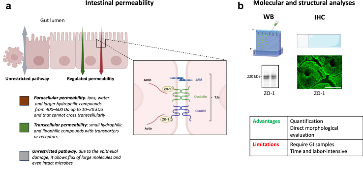
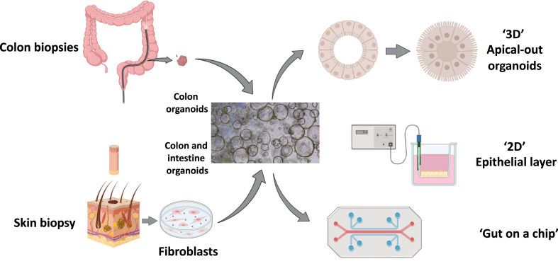
The intestinal epithelial barrier (IEB) plays a critical role in health and disease by regulating the absorption of nutrients, electrolytes and water while preventing gut translocation of pathogens. A compromised intestinal barrier has been reported in Parkinson's disease (PD) further reinforcing the assumption that PD is a gut-brain axis disorder and suggesting that gut-derived factors may participate in disease development and/or progression. However, the diversity of methodology between existing studies on gut permeability in PD, especially regarding the methods used for the evaluation of the IEB, has led to diverging results and it is definitely too early to draw any definite conclusions. We envision novel approaches, such as intestinal organoids and confocal laser endomicroscopy that could be used to study more precisely the IEB in PD.

Keywords: Confocal laser endomicroscopy; Intestinal epithelial barrier; Intestinal permeability; Organoids; Parkinson’s disease; Tight junctions; Ussing chamber.


Conflict of interest statement

Declaration of interests MS declares honorarium for scientific advisory board activities and lectures for Abbvie, Berlina, Biogen, Boston Scientific, Desitin, International Parkinson and Movement Disorders Society, Krka, Medtronic, Medis, Medison, Stada, TEVA and UCB and reports grants from Slovak Grant and Development Agency, Slovak Scientific Grant Agency, EU Renewal and Resilience Plan and European Regional Development Fund (ERDF) (all paid to the institution). PD reports grants from ANR (Agence nationale de la recherche) and Fondation pour la recherche sur le cerveau (Amadys), all paid to the institution. LLV reports grants from CHU de Nantes and France Parkinson (all paid to the institution). MRD reports grant from ANR (Agence nationale de la recherche), paid to the institution. KK reports funding from the Funding provided by the Slovak Scientific Grant Agency and the Slovak Research and Development (all paid to the institution).

Figures

Fig. 1
Fig. 1
Morphology and functions of the intestinal epithelial barrier (IEB). (a) Intestinal permeability is defined as the passage of molecules and ions from the gut lumen to the internal environment. There are three routes for such a passage: one non-specific due to a damaged epithelium (therefore called unrestricted pathways) and two specific routes, one across the plasma membrane of the epithelial cells (transcellular route) and the other one across protein networks called tight junctions (TJs) between two adjacent epithelial cells (paracellular route). Structurally, TJs are formed by a continuous mesh-like network of protein surrounding the apex of epithelial cells, which connects and tightens the space between adjacent cells. They consist of proteins including occludin, claudins, zonula occludens-1 (ZO-1) and junctional adhesion molecule (JAM) (b) the expression levels and the subcellular localisation of the TJs components can be analysed by western blot (WB) and immunohistochemistry (IHC). An example of western blot for the detection of ZO-1 in colonic biopsies (controls subjects), with a doublet migrating at 220 kDa (two isoforms) is shown. IHC performed in colonic mucosa (biopsy) from one control subject shows a typical distribution of ZO-1 with a honeycomb pattern (scale bar is 100 μm). Created in part with BioRender.com and with Servier Medical Art, licenced under the Creative Commons Attribution 3.0 Unported Licence.
Fig. 2
Fig. 2
Current techniques for the evaluation of intestinal permeability. IEB permeability is classically assessed using 3 main approaches: in vivo gut wall permeability tests, in vivo detection of translocation markers and ex vivo mucosal permeability tests with Ussing chambers. The advantages and limitations of each method are mentioned. Intestinal organoids and confocal laser endomicroscopy (CLE) are two promising additional approaches. Generated from biopsies, organoids allow the amplification of intestinal epithelial barrier in 3-dimensional structure and evaluation of intestinal epithelial barrier functions. The image shows human colonic organoids with a nuclear staining. CLE is a newly developed endoscopic technique that enables in vivo microscopic imaging of the gut mucosal layer together with intestinal permeability evaluated by fluorescein leakage. The image shows a colonic mucosa from a rat with chemically-induced colitis; fluorescein leakage is observed at the bottom of the crypts (white dots); scale bar is 20 μm. HPLC: high performance liquid chromatography. LC/MS: liquid chromatography–mass spectrometry. Created in part with BioRender.com and with Servier Medical Art, licenced under the Creative Commons Attribution 3.0 Unported Licence.
Fig. 3
Fig. 3
Potential use and application of human gut organoid technology for the evaluation of intestinal permeability. Intestinal organoids are derived from either induced pluripotent stem cells or colonic biopsies and form cyst-like structure. Epithelial permeability can be evaluated in 3-D spherical organoids. While these organoids form a closed hollow lumen, it is possible to reverse their polarity. The resulting “apical-out” organoids maintain a barrier function and provide an access to the luminal side. They allow barrier evaluation for example by measuring the passage of fluorescent probes and the expression levels of TJs genes and proteins. Alternatively, organoids could be cultured as 2-D monolayer in Transwell chambers. Permeability can be studied by measuring either transepithelial electrical resistance or flux of fluorescent markers. Eventually, organoids can be used in gut-on-chip models. These microfluidic devices are made of clear and flexible polymers into which several hollow channels or chambers are moulded. Created in part with BioRender.com and with Servier Medical Art, licenced under the Creative Commons Attribution 3.0 Unported Licence.

References

    1. Edwards L.L., Pfeiffer R.F., Quigley E.M., Hofman R., Balluff M. Gastrointestinal symptoms in Parkinson’s disease. Mov Disord. 1991;6:151–156. - PubMed
    1. Knudsen K., Fedorova T.D., Bekker A.C., et al. Objective colonic dysfunction is far more prevalent than subjective constipation in Parkinson’s disease: a colon transit and volume study. J Parkinsons Dis. 2017;7:359–367. - PubMed
    1. Savica R., Carlin J.M., Grossardt B.R., et al. Medical records documentation of constipation preceding Parkinson disease: a case-control study. Neurology. 2009;73:1752–1758. - PMC - PubMed
    1. Beach T.G., Adler C.H., Sue L.I., et al. Multi-organ distribution of phosphorylated alpha-synuclein histopathology in subjects with Lewy body disorders. Acta Neuropathol. 2010;119:689–702. - PMC - PubMed
    1. Gelpi E., Navarro-Otano J., Tolosa E., et al. Multiple organ involvement by alpha-synuclein pathology in Lewy body disorders. Mov Disord. 2014;29:1010–1018. - PubMed