. 2025 Jun 5;16(1):5207.
doi: 10.1038/s41467-025-60404-1.

Identification and characterisation of vaginal bacteria-glycan interactions implicated in reproductive tract health and pregnancy outcomes

Affiliations

Identification and characterisation of vaginal bacteria-glycan interactions implicated in reproductive tract health and pregnancy outcomes

Virginia Tajadura-Ortega et al. Nat Commun. .

Abstract

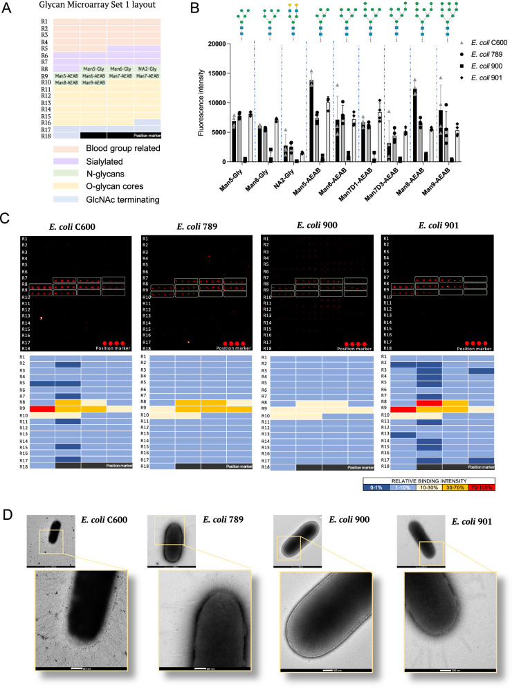
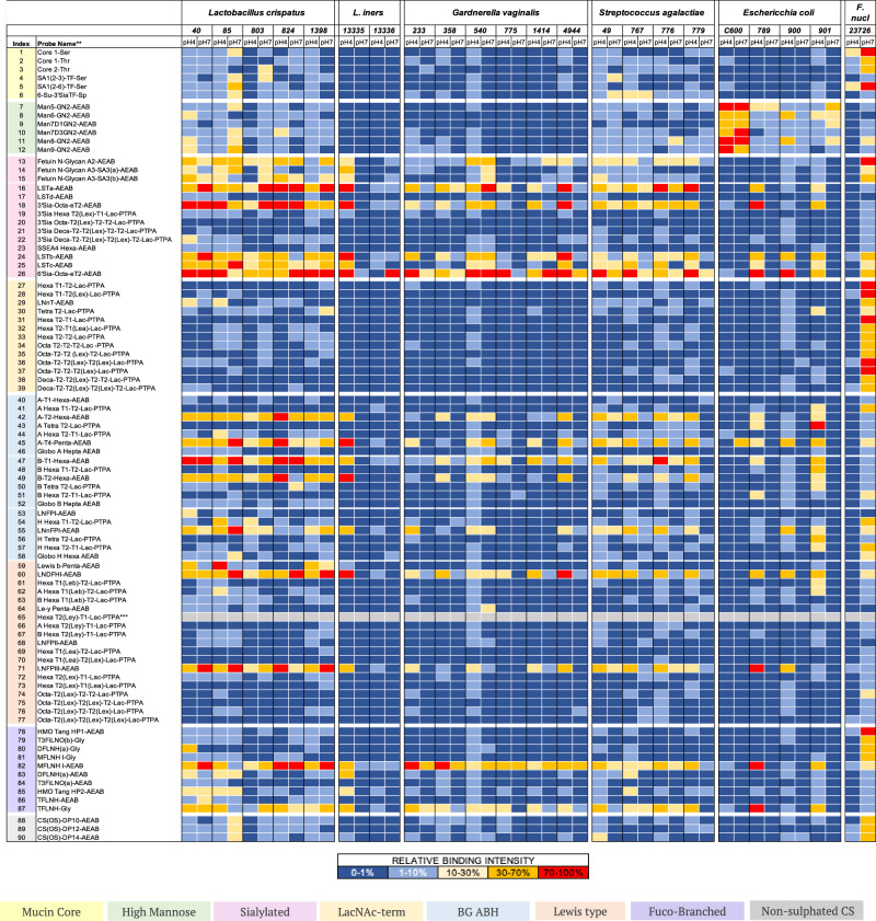
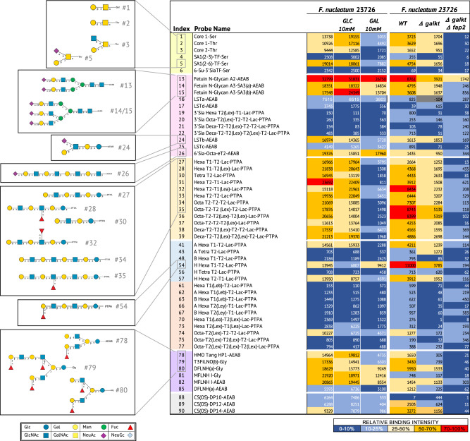
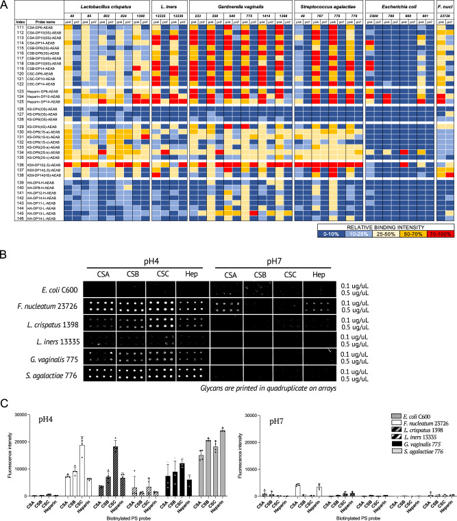
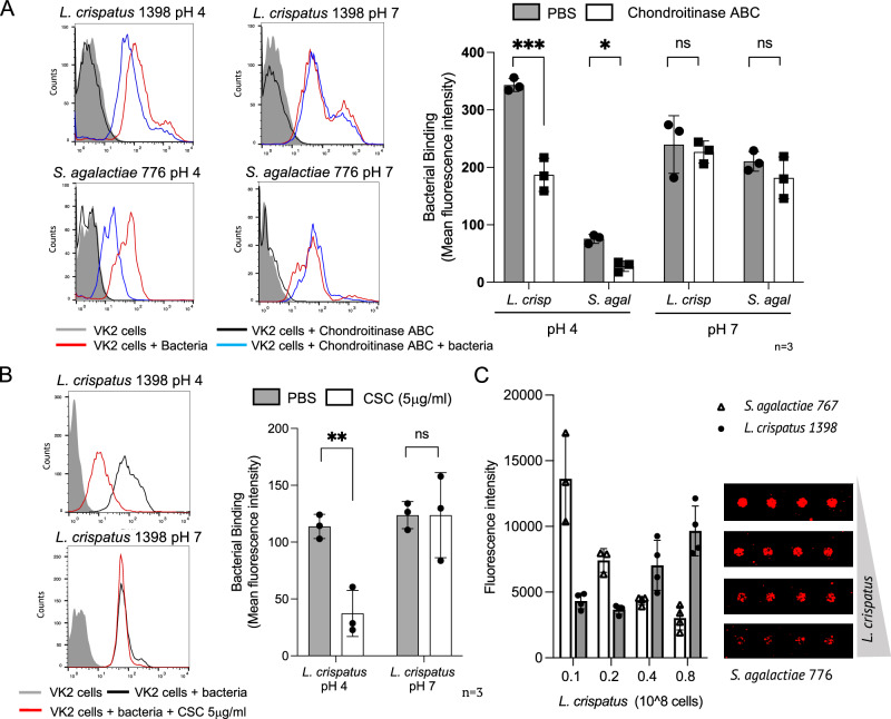
Lactobacillus displacement from the vaginal microbiome associates with adverse health outcomes and is linked to increased risk of preterm birth. Glycans mediate bacterial adhesion events involved in colonisation and infection. Using customised glycan microarrays, we establish glycan interaction profiles of vaginal bacteria implicated in reproductive health. Glycan binding signatures of the opportunistic pathogens Escherichia coli, Fusobacterium nucleatum and Streptococcus agalactiae to oligomannose N-glycans, galactose-terminating glycans and hyaluronic acid, respectively are highly distinct from Lactobacillus commensals. Binding to sulphated glycosaminoglycans by vaginal bacteria is pH dependent, as is binding to neutral and sialic acid-terminating glycans by F. nucleatum. Adhesion of Lactobacillus crispatus, Lactobacillus iners, Gardnerella vaginalis, S. agalactiae and F. nucleatum to vaginal epithelial cells is partially mediated by chondroitin sulphate. S. agalactiae binding to chondroitin sulphate C oligosaccharides is inhibited by L. crispatus. This study highlights glycans as mediators of vaginal bacterial binding events involved in reproductive health and disease.


Conflict of interest statement

Competing interests: The authors declare no competing interests.

Figures

Fig. 1
Fig. 1. Overview of the sequence-defined glycan library used in this study.
A Pie chart representing the glycan categories included in a customised sequence-defined glycan library for binding studies of vaginal microbiota. Numbers indicate the number of probes in main categories (Supplementary Data 1). Examples of glycans in each category are shown. PS polysaccharide, NS non-sulphated. B Diagram representing the methodology used for analysis of whole bacteria binding to sequence-defined glycans on microarrays. Glycans are covalently immobilised on NHS glass slides and binding performed with fluorescently labelled bacterial cultures of vaginal microbiota. C Bacterial collection with strain specification used for the study (Supplementary Data 4). * Strains purchased from ATCC or DMZ collections.
Fig. 2
Fig. 2. Comparison of glycan binding on glycan microarrays and electron microscopy of E. coli strains isolated from UTI patients.
A Printing layout of glycan Microarray set 1 indicating the location of the main glycan categories for the 69 sequence-defined glycan probes included in the microarray (Supplementary Data 1). B Bar charts showing the mean fluorescence intensities of bacterial binding signals to glycans printed on quadruplicated spots on the glycan Microarray set 1. Error bars represent the SD of quadruplicates for each glycan probe on the microarray (n = 1). Assay is representative of three independent experiments. Structures of bound glycans are shown. Fixed cells were used. Source data are provided as a Source Data File. C Top: Microarray images of fixed fluorescently labelled bacteria binding to glycans on Microarray set 1. Bottom: Heatmaps of relative binding strengths amongst the four E. coli strains tested to glycans on Microarray set 1. Colour code: Dark blue (0–1 %); Light blue (1–10 %); Yellow (10–30 %); Orange (30–70%); Red (70–100%). 100%, the maximum binding score observed across the four experiments. D Electron-microscopy images of whole bacterial cells of E. coli strains C600, 789, 900, and 901. Enlargements showing differences in fimbriae sizes. White bars in enlargements indicate 200 nm. Images are representative of two independent experiments.
Fig. 3
Fig. 3. Binding of bacteria from the vaginal microbiota to neutral and sialic acid terminating glycans on glycan microarrays.
Heatmap showing the relative binding intensities of live fluorescently labelled bacterial cultures of L. crispatus, L. iners, G. vaginalis, S. agalactiae, and fixed E. coli and F. nucleatum strains to 90 neutral and sialic acid terminating glycans at pH 4 and pH 7 on Microarray set 2 with glycans printed at high density (300 μM). Colour code: Dark blue (0–1%); Light blue (1–10%); Yellow (10–30%); Orange (30–70%); Red (70–100%). 100%, the maximum binding score observed across pH 4 and pH 7 for a given strain. Glycan probes are grouped according to their sequences into categories and annotated by the coloured panels defined at the bottom of the figure. Histograms of representative strains are shown in Supplementary Fig. 3. Grey cells correspond to glycan probe #65 that did not pass quality control in this set.
Fig. 4
Fig. 4. Glycan binding studies of wt and Δfap2 Fusobacterium nucleatum with sequence-defined covalent microarrays reveals binding to sialylated, fucosylated and galactose-terminating glycans.
(Left panels) Heatmaps of relative binding strengths of fixed fluorescently labelled F. nucleatum 23726 to neutral and sialylated glycans at pH7 in the presence of 10 mM galactose, 10 mM glucose or buffer only. Source data are provided in Source Data File 5. (Right panels) Heatmaps of relative binding strengths of fixed fluorescently labelled wt, Δgalkt and Δgalkt Δfap2 F. nucleatum 23726 to neutral and sialylated glycans at pH7. Values on heatmap indicate the mean fluorescence intensities of quadruplicate spots on Microarray set 2a and 2b. Colour code for binding intensities: Dark blue (0–10%); Light blue (10–25%); Yellow (25–50%); Orange (50–70%); Red (70–100%). 100%, the maximum binding score observed across the three conditions tested. Glycan structures corresponding to selected bound glycan probes are shown. Glycans categories are coloured as follows: Yellow: Mucin core related; Pink: Sialylated; Orange: LacNAc terminating; Blue: Blood group A, B, H glycans; Peach: Lewis blood group related; Purple: Fucosylated branched glycans from human milk; Grey: Non-sulphated chondroitin sulphate. * (-104) Grey cells were flagged for artefact/background on slide.
Fig. 5
Fig. 5. Glycosaminoglycan binding by bacteria of the vaginal microbiota.
A Heatmap showing the relative binding intensities to 36 GAG oligosaccharides at pH4 and pH7 of fixed fluorescently labelled strains of E. coli and F. nucleatum, and live L. crispatus, L. iners, G. vaginalis and S. agalactiae. The average rank of the mean fluorescence intensities from at least two independent experiments on Microarray set 5 was calculated (Source Data file 6) and the resulting values coloured as follows: Dark blue (0–10 %); Light blue (10–25 %); Yellow (25–50 %); Orange (50–70%); Red (70–100%). 100%, the maximum binding score observed across pH4 and pH7 for a given strain. B Glycan microarray images of fluorescently labelled E. coli C600, F. nucleatum 23726, L. crispatus 1398, L. iners 13335, G. vaginalis 775 and S. agalactiae 776 binding to GAG polysaccharides at pH4 or pH7. Images are representative of three independent experiments. C Bar charts showing the mean fluorescence intensities of bacterial binding at pH4 or pH7 to GAG polysaccharides on Microarray set 6 printed at 0.5 mg/ml. Error bars represent SD of quadruplicate spots for each polysaccharide on the microarray (n = 1). Assay is representative of three independent experiments. Source data are provided in Source Data File 7.
Fig. 6
Fig. 6. Analysis of bacterial binding to VK2 vaginal epithelial cells and glycan binding competition by bacteria on glycan microarrays.
A Flow cytometry-based analysis of live bacterial binding to VK2 cells treated or not with chondroitinase ABC. Bar charts show the mean fluorescence intensities of the bound bacteria at pH4 and pH7. Error bars represent SD of three independent experiments for each condition (n = 3). B Flow cytometry-based analysis of the binding of L. crispatus preincubated or not with CSC polysaccharide to VK2 cells. Bar charts show the mean fluorescence intensities of the bound bacteria at pH4 and pH7. Error bars represent SD of three independent experiments for each condition (n = 3). C Left: Bar charts show the mean fluorescence intensities of the binding at pH4 of fluorescently labelled S. agalactiae 776 and L. crispatus 1398 to a CSC 14-mer oligosaccharide in the presence of increasing amounts of L. crispatus 1398. Error bars represent SD of triplicates on the microarray for each glycan (n = 1). One experiment representative of two independent experiments is shown. Right: Representative images of S. agalactiae 776 cells binding to CSC 14-mer on the microarray in the presence of increasing amounts of L. crispatus 1398. *** p < 0.0001; *p < 0.039; ns: non-significant; 2-way ANOVA (A) and **p < 0.0067; ns: non-significant B). Source data are provided in the Source Data File 7.

Similar articles

References

    1. Golob, J. et al. Microbiome Preterm Birth DREAM Challenge: Crowdsourcing machine learning approaches to advance preterm birth research. Cell Rep. Med.5, 101350 (2024). - PMC - PubMed
    1. Huang, C. et al. Meta-analysis reveals the vaginal microbiome is a better predictor of earlier than later preterm birth. BMC Biol.21, 199 (2023). - PMC - PubMed
    1. Gudnadottir, U. et al. The vaginal microbiome and the risk of preterm birth: a systematic review and network meta-analysis. Sci. Rep.12, 7926 (2022). - PMC - PubMed
    1. van de Wijgert, J. & Jespers, V. The global health impact of vaginal dysbiosis. Res Microbiol168, 859–864 (2017). - PubMed
    1. Callahan, B. et al. Replication and refinement of a vaginal microbial signature of preterm birth in two racially distinct cohorts of US women. Proc. Natl. Acad. Sci. USA114, 9966–9971 (2017). - PMC - PubMed

LinkOut - more resources