

Tendon modification with percutaneous Ultrasound-Guided Tenotomy using TENEX®: A histological and macroscopic analysis of a bovine cadaveric model
- PMID: 40469069
- PMCID: PMC12136825
- DOI: 10.1016/j.inpm.2025.100590
Tendon modification with percutaneous Ultrasound-Guided Tenotomy using TENEX®: A histological and macroscopic analysis of a bovine cadaveric model
Abstract
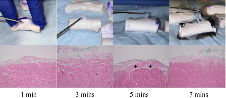
Background: Chronic tendinopathy treatment remains elusive; however, percutaneous Ultrasound-Guided Tenotomy using TENEX® (PUTT) has demonstrated promising clinical outcomes, but the mechanism of action is not clearly defined. This study aims to describe potential mechanisms using an ex-vivo animal model. The objective of the study is to examine the histological effects of PUTT on the bovine soleus tendon across varying treatment durations.
Methods: Twelve bovine soleus tendons were allocated to four cohorts to undergo PUTT for 1, 3, 5, and 7 min. Each specimen was treated on one side, the opposite side serving as a control. Macroscopic and microscopic analyses were conducted to assess tendon sheath, fascicle, and perineural disruption. Fascicle penetration was measured using ImageJ software. Statistical analyses were performed using Analysis of Variance (ANOVA), with significance levels set at p < 0.05.
Results: Macroscopic and microscopic examination revealed progressive separation of the paratenon, peritendinous nerves, and fascicles, correlating with increased treatment duration. Fascicle penetration depths were 0.1 mm, 2.57 mm, 2.61 mm, and 3.93 mm at 1, 3, 5, and 7 min, respectively. ANOVA confirmed significant differences among groups (F (3, 8) = 620.898, p < 0.001), with a large effect size (η2 = 0.996. Tukey's Honest Significant Difference (HSD) test revealed significant differences between most groups (p < 0.001), except between the 3-min and 5-min treatments, which showed no significant difference (p = 0.969).
Conclusion: PUTT induces significant structural changes in the paratenon and fascicle layer with longer treatment duration, resulting in more pronounced modifications.
Keywords: Chronic; Future; Nociceptors; Pain; Paratenon; Tendinopathy.
© 2025 The Authors.
Conflict of interest statement
Authors provided Conflict of Interest forms for all authors.
Figures


References
-
- Irby A., Gutierrez J., Chamberlin C., Thomas S.J., Rosen A.B. Clinical management of tendinopathy: a systematic review of systematic reviews evaluating the effectiveness of tendinopathy treatments. Scand J Med Sci Sports. 2020;30(10):1810–1826. - PubMed
-
- Alfredson H. Chronic midportion Achilles tendinopathy: an update on research and treatment. Clin Sports Med. 2003;22(4):727–741. - PubMed
-
- Battista C.T., Dorweiler M.A., Fisher M.L., Morrey B.F., Noyes M.P. Ultrasonic percutaneous tenotomy of common extensor tendons for recalcitrant lateral epicondylitis. Tech Hand Up Extrem Surg. 2018;22(1):15–18. - PubMed
-
- Burke C.J., Adler R.S. Ultrasound-guided percutaneous tendon treatments. AJR Am J Roentgenol. 2016;207(3):495–506. [published correction appears in AJR Am J roentgenol. 2016 sep; 207(3):689. Doi: 10.2214/AJR.16.16934] - PubMed
LinkOut - more resources
Full Text Sources
