. 2025 May 21:12:1509086.
doi: 10.3389/fnut.2025.1509086. eCollection 2025.

Coptis japonica Makino ethanol extracts attenuates cancer cachexia induced muscle and fat wasting through inhibition of the STAT3 signaling pathway

Affiliations

Coptis japonica Makino ethanol extracts attenuates cancer cachexia induced muscle and fat wasting through inhibition of the STAT3 signaling pathway

Yo Sep Hwang et al. Front Nutr. .

Abstract

Cancer cachexia is a complex syndrome marked by appetite loss, weakness, fatigue, significant weight loss, and depletion of both adipose and muscle tissue, driven by metabolic and inflammatory alterations caused by tumors. Cachexia is a critical contributor to poor cancer prognosis, often leading to reduced efficacy of treatments. Coptis japonica Makino (CJM) is a medicinal herb widely used in Asia, known for its anti-inflammatory and metabolic regulatory properties. However, its potential role in cancer cachexia has not yet been explored. This research aimed to explore the potential of CJM extracts (CJME) in mitigating cancer cachexia in both myotubes treated with CT26 conditioned medium (CM) and in a CT26-induced cancer cachexia mouse model. Our results demonstrated that CJME significantly decreased the mRNA and protein levels of muscle-specific E3 ubiquitin ligases Atrogin-1 and MuRF1 in myotubes exposed to CT26 CM. Furthermore, CJME notably enhanced the protein levels of myosin heavy chain (MyHC). In the mouse model of CT26-induced cancer cachexia, severe loss of muscle and fat was observed. however, CJME effectively countered this wasting and restored abnormal biochemical parameters such as CK, albumin, triglycerides (TG), cholesterol, high-density lipoprotein (HDL), and low-density lipoprotein (LDL) associated with cancer cachexia. Moreover, CJME reduced interleukin-6 (IL-6) levels in both CT26 CM-stimulated myotubes and the serum of CT26-induced cancer cachexia mice. The mechanism underlying these effects appears to involve the suppression of STAT3 activation by CJME. These findings suggest that CJME has potential as a therapeutic candidate in the management of cancer cachexia.

Keywords: Coptis japonica Makino; IL-6; STAT3; cancer cachexia; muscle atrophy.


Conflict of interest statement

The authors declare that the research was conducted in the absence of any commercial or financial relationships that could be construed as a potential conflict of interest. The author(s) declared that they were an editorial board member of Frontiers, at the time of submission. This had no impact on the peer review process and the final decision.

Figures
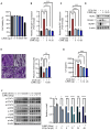
Figure 1
Figure 1
The effects of CJME on CT26 conditioned medium (CM) induced-myotube atrophy. The C2C12 myoblasts were differentiated for 2 days and induced to form myotubes. (A) Effect of CJME on differentiated myotubes viability were determined using WST-1 assay. (B) Indicated CJME concentrations were used for pretreatment 1 h prior to treatment with CT26 CM for 48 h in differentiated myotubes. mRNA levels of (B) Atrogin-1 and (C) MuRF1 were determined using real-time PCR. Relative mRNA expression levels were normalized to those of GAPDH. (D) Protein levels of MyHC, Atrogin-1 and MuRF1 were analyzed via immunoblotting in cells cultured as described in B. (E) Differentiated myotubes were cultured as described in B, fixed with 4% PAF and stained with Crystal violet. Representative images of stained myotubes were shown (left). The scale bar represents 200 μm. Myotube thickness was measured using Image J 1.53 software (right) (F) Differentiated myotubes were pretreated with 20 μg/mL of CJME for 1 h and exposed to CT26 CM for various times (0, 3, 6, 12, 24, and 48 h). Then, phosphorylation of STAT1, STAT2, STAT3, ERK, JNK, p38, IKKα/β and IκBα was examined using immunoblotting (left). Quantified data for STAT3 were measured using Image J 1.53 software (right) (G) Differentiated myotubes were cultured as described in B, secreted levels of IL-6 in the culture medium were measured using ELISA. Data are representative of either two independent experiments. Data are presented in terms of the mean ± standard deviation and analyzed using a one-wayANOVA or Student's t-test. P value of <0.05 (*), <0.01 (**), <0.001 (***), or <0.0001 (****) were considered statistically significant.
Figure 2
Figure 2
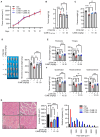
The effects of CJME on muscle wasting in CT26-induced cancer cachexia model. CT26 cells (2.5 × 10⁶/mouse) were subcutaneously injected into the flank of BALB/c mice, and starting from day 7 post-injection, CJME (10 and 20 mg/kg) was orally administered every 2 days for 3 weeks. The negative control group (Ctrl) was not injected with CT26 cells, and the positive control group (CT26) was orally administered PBS at the same dose as CJME. (A) Tumor volume was measured every 3 days starting from 7 days after the injection of CT26 cells. (B) After 4 weeks of CT26 cell injection, the tumors were excised, and tumor weight and (C) Carcass weight without the tumor mass were measured. (D) Representative images of the hind limb (Gastrocnemius, Soleus, Tibialis anterior muscle complex) were shown (left), hind limb muscles weight was measured in CT26-induced cachexia mice model (right). (E) Pectoralis and triceps muscle weight was measured in CT26-induced cachexia mice model. (F) Quadriceps, tibialis anterior (TA) and gastrocnemius muscle weight was measured in CT26-induced cachexia mice model. (G) Gastrocnemius muscle were stained with H&E staining, and representative images were shown (left). The scale bar represents 50 μm. The average cross-sectional area (CSA) of gastrocnemius muscle fiber was quantified by Image J 1.53 software (middle) and fiber frequency distribution was quantified by Image J 1.53 software (right). (n = 4 mice per group). Data are representative of either two independent experiments. Data are presented in terms of the mean ± standard deviation and analyzed using a one-wayANOVA or Student's t-test. P < 0.05 (*), <0.01 (**), <0.001 (***), or <0.0001 (****) were considered statistically significant.
Figure 3
Figure 3
The effects of CJME on fat wasting in CT26-induced cancer cachexia model. CT26 cells (2.5 × 10⁶/mouse) were subcutaneously injected into the flank of BALB/c mice, and starting from day 7 post-injection, CJME (10 and 20 mg/kg) was orally administered every 2 days for 4 weeks. The negative control group (Ctrl) was not injected with CT26 cells, and the positive control group (CT26) was orally administered PBS at the same dose as CJME. (A) Representative images of the eWAT were shown (left), eWAT weight was measured (right). (n = 4 mice per group). (B, C) ingWAT and iBAT weight was measured. (D) eWAT was stained with H&E staining, and representative images were shown (left). The scale bar represents 50 μm. The average cross-sectional area (CSA) of eWAT was quantified by Image J 1.53 software (middle) and frequency distribution of adipocyte cell area was quantified by Image J 1.53 software (right). (n = 4 mice per group). Data are representative of either two independent experiments. Data are presented in terms of the mean ± standard deviation and analyzed using a one-wayANOVA or Student's t-test. P < 0.01 (**), <0.001 (***), or <0.0001 (****) were considered statistically significant.
Figure 4
Figure 4
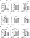
The effect of CJME on biochemical parameters and pro-inflammatory cytokines level in serum of CT26 induced-cancer cachexia model. CT26 cells (2.5 × 10⁶/mouse) were subcutaneously injected into the flank of BALB/c mice, and starting from day 7 post-injection, CJME (10 and 20 mg/kg) was orally administered daily for 1 weeks. The negative control group (Ctrl) was not injected with CT26 cells, and the positive control group (CT26) was orally administered PBS at the same dose as CJME. (A–F) Creatine kinase (CK) (A), albumin (B), triglycerides (TG) (C), cholesterol (D), high-density lipoprotein (HDL) (E) and low-density lipoprotein (LDL, F) levels were measured in serum of CT26 induced mice. (G–I) IL-6 (G), IL-1β (H) and TNF-α (I) levels were measured in serum of CT26 induced mice (n = 4 mice per group). Data are representative of either two independent experiments. Data are presented in terms of the mean ± standard deviation and analyzed using a one-wayANOVA or Student's t-test. P < 0.05 (*), <0.01 (**), <0.001 (***), or <0.0001 (****) were considered statistically significant.
Figure 5
Figure 5
LC-MS profiles of the 70% EtOH extracts of Coptis japonica Makino. Coptisine (1), Palmatine (2), and Berberine (3).

Similar articles

References

    1. Ni J, Zhang L. Cancer cachexia: definition, staging, and emerging treatments. Cancer Manag Res. (2020) 12:5597–605. 10.2147/CMAR.S261585 - DOI - PMC - PubMed
    1. Baracos VE, Martin L, Korc M, Guttridge DC, Fearon KCH. Cancer-associated cachexia. Nat Rev Dis Primers. (2018) 4:17105. 10.1038/nrdp.2017.105 - DOI - PubMed
    1. Kasprzak, A. The role of tumor microenvironment cells in colorectal cancer (CRC) cachexia. Int J Mol Sci. (2021) 22:1565. 10.3390/ijms22041565 - DOI - PMC - PubMed
    1. Argiles JM, Busquets S, Stemmler B, Lopez-Soriano FJ. Cancer cachexia: understanding the molecular basis. Nat Rev Cancer. (2014) 14:754–62. 10.1038/nrc3829 - DOI - PubMed
    1. Rom O, Reznick AZ. The role of E3 ubiquitin-ligases MuRF-1 and MAFbx in loss of skeletal muscle mass. Free Radic Biol Med. (2016) 98:218–30. 10.1016/j.freeradbiomed.2015.12.031 - DOI - PubMed

LinkOut - more resources