. 2025 May 21:16:1572117.
doi: 10.3389/fmicb.2025.1572117. eCollection 2025.

Functional loss of rffG and rfbB, encoding dTDP-glucose 4,6-dehydratase, alters colony morphology, cell shape, motility and virulence in Salmonella Typhimurium

Affiliations

Functional loss of rffG and rfbB, encoding dTDP-glucose 4,6-dehydratase, alters colony morphology, cell shape, motility and virulence in Salmonella Typhimurium

Subhashish Chakraborty et al. Front Microbiol. .

Abstract

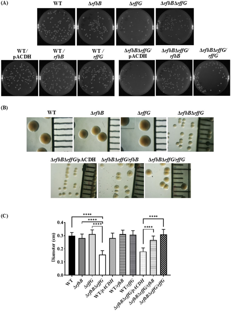
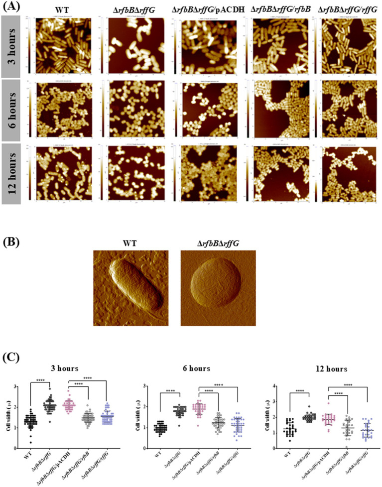
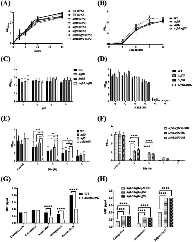
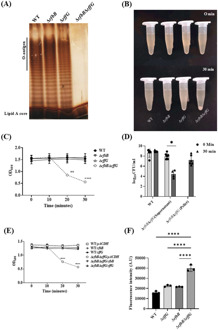
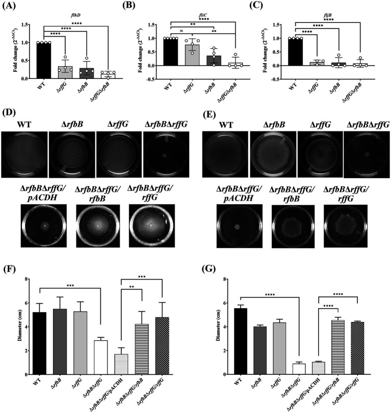
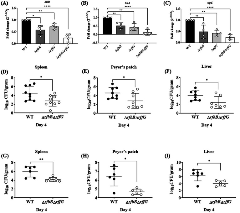
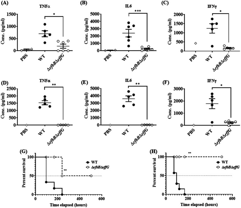
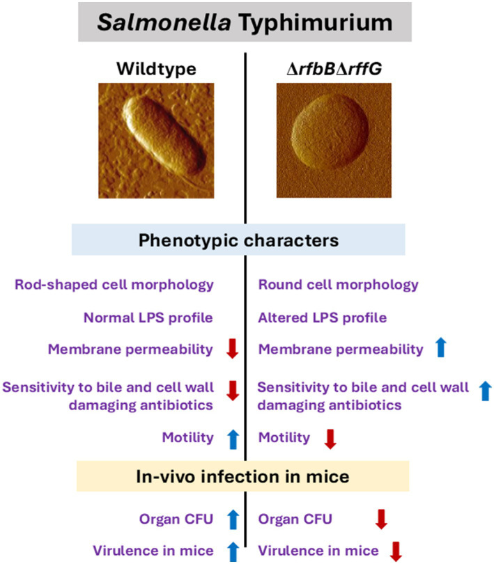
Lipopolysaccharide (LPS) O-antigen and enterobacterial common antigen (ECA) play crucial roles in maintaining the outer membrane in Gram-negative bacteria. Mutations in the biosynthetic pathways of LPS and ECA may lead to accumulation of intermediates, resulting in morphological changes and activation of stress responses. However, the functional consequences of abrogation of both O-antigen and ECA synthesis in Salmonella enterica serovar Typhimurium (S. Typhimurium) are not well investigated. In this study, we generated single and double-deletion mutants of rfbB and rffG, encoding dTDP-glucose 4,6-dehydratase paralogs that are important in the synthesis of both O-antigen and ECA. Importantly, mutations in the dTDP-D-glucose 4,6-dehydratase encoding gene in humans are known to cause Catel-Manzke syndrome, a rare genetic disease. All four strains, i.e., wild type (WT), ΔrfbB, ΔrffG and ΔrfbBΔrffG, grew well in rich Luria Bertani (LB) liquid media at 37°C; however, the functional loss of both rfbB and rffG, but not in single-deletion strains, resulted in round cell morphology and smaller colony size in LB agar plates. There was no significant differences in the growth of the four strains in minimal media at 37°C (nutritional deficiency), in LB at 42°C (high temperature), acidic pH or LB with 3-4% NaCl (high osmolarity; however the ΔrfbBΔrffG strain was hypersensitive to bile and cell wall-targeting antibiotics). These results demonstrated that the ΔrfbBΔrffG strain was sensitive to some stress conditions. Interestingly, the ΔrfbBΔrffG strain displayed an altered LPS profile, autoaggregated rapidly compared to the WT and the single mutant strains and showed high N-phenylnaphthylamine (NPN) fluorescence indicating greater surface hydrophobicity. Furthermore, transcriptomic analysis identified flagellar and SPI-1 pathways to be highly downregulated in ΔrfbBΔrffG which led to impaired swimming as well as swarming motility, lower adhesion and invasion of HeLa cells. Importantly, the ΔrfbBΔrffG strain was less proficient in colonizing Peyer's patches, spleen and liver, was unable to induce pro-inflammatory cytokines and was attenuated in both the oral and intraperitoneal models of S. Typhimurium infection in mice. Overall, this study highlights the importance of rfbB and rffG in maintaining cell wall and cell membrane integrity, colony and cellular morphology, motility and virulence in S. Typhimurium.

Keywords: 6-dehydratase; Salmonella Typhimurium; dTDP-D-glucose 4; enterobacterial common antigen; lipopolysaccharide; rfbB; rffG.


Conflict of interest statement

The authors declare that the research was conducted in the absence of any commercial or financial relationships that could be construed as a potential conflict of interest.

Figures

Figure 1
Figure 1
Salmonella Typhimurium ΔrfbBΔrffG strain forms smaller colonies on LB agar. (A) S. Typhimurium WT, ΔrfbB, ΔrffG, ΔrfbBΔrffG and complemented strains were plated on LB agar plates, and their images were captured after 16 h of incubation at 37°C. (B) Representative images depicting individual colony morphology of the above-mentioned strains obtained through a light microscope. (C) Diameter of the colony of WT, ΔrfbB, ΔrffG and ΔrfbBΔrffG and the complemented strains on LB agar plates. Statistical analysis was performed using one-way ANOVA, where ****p < 0.0001. Data are representative of three independent experiments plotted as mean ± SEM. Scale Bar for (B): 1 mm per unit.
Figure 2
Figure 2
Complementation of S. Typhimurium ΔrfbBΔrffG strain with WT copy of rfbB or rffG restores cellular width. (A) The bacterial strains were grown for the indicated time points (3, 6 or 12 h) and AFM images were acquired in the non-contact mode. (B) Single cell images of the WT and the ΔrfbBΔrffG at 6 h. (C) Quantification of the cellular width was performed and for each condition, the width of at least 50 cells was determined. Data are representative of 3 independent experiments plotted as mean ± SEM. Statistical analysis was performed using two-way ANOVA, where * p < 0.05; ** p < 0.01; *** p < 0.001 and **** p < 0.0001.
Figure 3
Figure 3
Salmonella Typhimurium ΔrfbBΔrffG strain is susceptible to bile and cell wall targeting antibiotics. S. Typhimurium WT and ΔrfbBΔrffG were grown (A) in LB at 37°C and 42°C (B) in minimal media. To test the effects of growth at high temperature, S. Typhimurium WT, ΔrfbB, ΔrffG, and ΔrfbBΔrffG were grown in LB and media at 37°C and 42°C overnight. Post incubation, the cultures were normalized to an optical density (OD) of 2. Subsequently, 0.2% (v/v) of normalized cultures were inoculated into LB medium and incubated for 3 h. Following this, 0.2% (v/v) of the culture was transferred to respective tubes containing LB media and incubated at 37°C and 42°C with shaking at 160 RPM. Optical density measurements were recorded at specified time points. S. Typhimurium WT, ΔrfbB, ΔrffG and ΔrfbBΔrffG growth in 5 mL LB broth (C) of different pH, and (D) with different concentrations of NaCl. (E) S. Typhimurium WT, ΔrfbB, ΔrffG and ΔrfbBΔrffG and (F) complemented strains, ΔrfbBΔrffG/pACDH, ΔrfbBΔrffG/rfbB, ΔrfbBΔrffG/rffG strains were treated with the indicated concentrations of bile and grown for 8 h at 37°C and 160 rpm. Bacterial growth was quantified by measuring OD at 600 nm. Epsilometer Test (E-Test) results to determine MIC of the (G) WT and the ΔrfbBΔrffG strains as well as the (H) complemented strains. Briefly, overnight grown cultures were normalized to OD 2 at 600 nm and plated onto the Mueller Hinton agar plates by spread plate method. Pre-coated Ezy-MIC strips with selected antibiotics were placed in the middle of the agar plates with the help of a sterile swab and the plates were incubated at 37°C for 18 h. Data are representative of three independent experiments and plotted as mean ± SEM. Statistical analysis was performed using two-way ANOVA, where * p < 0.05; ** p < 0.01; *** p < 0.001 and **** p < 0.0001.
Figure 4
Figure 4
Salmonella Typhimurium ΔrfbBΔrffG strain displays a truncated O-antigen profile, rapid auto-aggregation and binds more to NPN dye. (A) S. Typhimurium WT, ΔrfbB, ΔrffG and ΔrfbBΔrffG strains were grown in LB broth at 37°C for 15 h. LPS was isolated and resolved on a 12% SDS PAGE. (B,C) Auto-aggregation behavior of the WT and the gene-deleted strains. Briefly, 1 mL of 1.5 OD normalized, S. Typhimurium WT, ΔrfbB, ΔrffG and ΔrfbBΔrffG cultures in LB broth were kept in an upright position for 30 min. At every 10-min interval, 100 μL was aspirated from the top of the solution, and the OD was measured at 600 nm. (D) Post 30 min, the supernatant was aspirated from the top of the culture, and the residual pellet was resuspended in 1 mL of sterile LB. Appropriate dilutions were then plated, and colony-forming units (CFU) were recorded. (E) Complementation with the WT copy of the gene(s) show a rescue in the phenotype. (F) S. Typhimurium ΔrfbBΔrffG accumulates higher amounts of the NPN dye. Briefly, the representative bacterial strains were grown to an OD of 0.5 at 600 nm. Cells were harvested, and the bacterial pellet was washed with 5 mM HEPES (pH 7.2) and adjusted to an OD of 0.5 at 600 nm. NPN was added to each well at a concentration of 10 μM. Fluorescence excitation and emission were measured at 350 nm and 420 nm, respectively. Statistical analysis for this assay was performed using one-way ANOVA, where ** p < 0.01. Data are representative of three independent experiments and plotted as mean ± SEM. Statistical analysis was performed using two-way ANOVA, where ** p < 0.01 and **** p < 0.0001.
Figure 5
Figure 5
Salmonella Typhimurium ΔrfbBΔrffG strain is compromised in swimming and swarming motility. Total RNA was extracted from the bacterial samples after 3 h of growth in LB broth at 37°C and 160 rpm. A qRT-PCR analysis was performed to monitor the expression of the genes, (A) flhD, (B) fliC and (C) fljB and the expression normalized with the reference gene, gmk. The (D) swimming and (E) swarming motility assays were performed on 0.3 and 0.5% agar, respectively. Equal amounts of cultures were inoculated at the center of the motility-agar plates and the plates were incubated at 37°C for 8 h in an upright condition. The distance covered by each strain after 8 h post inoculation for (F) swimming and (G) swarming motility was measured and analyzed using ImageJ software. Multiple measurements were obtained for each strain. Data are representative of three independent experiments and plotted as mean ± SEM. Statistical analysis was performed using one-way ANOVA, where * p < 0.05; ** p < 0.01; *** p < 0.001 and **** p < 0.0001.
Figure 6
Figure 6
Salmonella Typhimurium ΔrfbBΔrffG strain is less proficient in colonizing different organs. Total RNA was extracted from the bacterial samples after 3 h of growth in LB broth at 37°C and 160 rpm. A qRT-PCR analysis was performed to monitor the expression of the SPI-1 genes, (A) hilD (B) hilA and (C) sipC, and the expression normalized with the reference gene, gmk. Bacterial burden in the organs was estimated after (D–F) oral and (G-I) intraperitoneal infection of C57BL/6 mice with the WT and the ΔrfbBΔrffG strains. Infected mice were sacrificed on day 4 post infection, organs were harvested, and the bacterial burden in the spleen, Peyer’s Patch and the liver was estimated by plating serial dilutions of the tissue homogenate on LB agar plate. Gene expression analysis data are representative of three independent experiments plotted as mean ± SEM and statistical analysis was performed using one-way ANOVA, where * p < 0.05; ** p < 0.01; *** p < 0.001 and **** p < 0.0001. For estimating bacterial burden in organs, statistical analysis was performed using the Mann Whitney U test.
Figure 7
Figure 7
Salmonella Typhimurium ΔrfbBΔrffG strain is highly attenuated in oral and intraperitoneal mice models of infection. The levels of pro-inflammatory cytokines (A) TNFα (B) IL6, and (C) IFNγ at day 4 post infection were estimated from the serum collected from mice infected orally with 1×10⁸ CFU of the WT and ΔrfbBΔrffG strains. Similarly, mice infected intraperitoneally with 1×10³ CFU of S. Typhimurium WT and ΔrfbBΔrffG were sacrificed at day 4 post infection and the serum levels of cytokines, (D) TNFα (E) IL6 and (F) IFNγ were quantified. Kaplan–Meier survival analysis comparing mice survival for 21 days upon infection with the WT and the ΔrfbBΔrffG strains, infected either, (G) orally or (H) intraperitoneally. Data are representative of three independent experiments plotted as mean ± SEM. Statistical analysis was performed using Log-rank (Mantel-Cox) test, where **p < 0.005 and ***p < 0.0001. For cytokine estimation, statistical analysis was performed using Kruskal Wallis test, where *p < 0.05; **p < 0.01 and ***p < 0.001.
Figure 8
Figure 8
Salmonella Typhimurium WT and ∆rfbBrffG show distinct physiological and phenotypic differences. The genes, rfbB and rffG, encode the enzyme dTDP-glucose 4,6- dehydratase, which catalyzes intermittent steps of O-antigen and ECA biosynthesis. Functional loss of dTDP-glucose 4,6-dehydratase renders S. Typhimurium strains incapable of synthesizing both O-antigen and ECA. This leads to profound physiological differences between the S. Typhimurium, WT and the ΔrfbBΔrffG strains. The ΔrfbBΔrffG strain exhibits distinct cellular morphology, altered LPS profile, increased outer membrane permeability and susceptibility to bile and cell wall targeting antibiotics. Functional loss of dTDP-glucose 4,6-dehydratase also led to inhibition of motility and a reduced ability to colonize different organs in the mouse model of infection. Consequently, the ΔrfbBΔrffG strain displayed significant virulence attenuation as compared to the WT.

Similar articles

References

    1. Allard S. T. M., Beis K., Giraud M.-F., Hegeman A. D., Gross J. W., Wilmouth R. C., et al. . (2002). Toward a structural understanding of the dehydratase mechanism. Structure 10, 81–92. doi: 10.1016/S0969-2126(01)00694-3, PMID: - DOI - PubMed
    1. Allard S. T. M., Giraud M.-F., Whitfield C., Graninger M., Messner P., Naismith J. H. (2001). The crystal structure of dTDP-d-glucose 4,6-dehydratase (RmlB) from Salmonella enterica serovar typhimurium, the second enzyme in the dTDP-l-rhamnose pathway. J. Mol. Biol. 307, 283–295. doi: 10.1006/jmbi.2000.4470, PMID: - DOI - PubMed
    1. Beis K., Allard S. T. M., Hegeman A. D., Murshudov G., Philp D., Naismith J. H. (2003). The structure of NADH in the enzyme dTDP-d-glucose dehydratase (RmlB). J. Am. Chem. Soc. 125, 11872–11878. doi: 10.1021/ja035796r, PMID: - DOI - PubMed
    1. Bhaskarla C., Das M., Verma T., Kumar A., Mahadevan S., Nandi D. (2016). Roles of Lon protease and its substrate MarA during sodium salicylate-mediated growth reduction and antibiotic resistance in Escherichia coli. Microbiology 162, 764–776. doi: 10.1099/mic.0.000271, PMID: - DOI - PubMed
    1. Bode N. J., Debnath I., Kuan L., Schulfer A., Ty M., Pearson M. M. (2015). Transcriptional analysis of the MrpJ network: modulation of diverse virulence-associated genes and direct regulation of mrp fimbrial and flhDC flagellar operons in Proteus mirabilis. Infect Immun. 83:2542–56. - PMC - PubMed

LinkOut - more resources