

Characterization of extracellular vesicles and miRNA released by cerebral organoids
- PMID: 40469891
- PMCID: PMC12133712
- DOI: 10.1016/j.crtox.2025.100229
Characterization of extracellular vesicles and miRNA released by cerebral organoids
Abstract
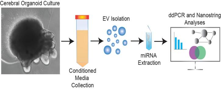
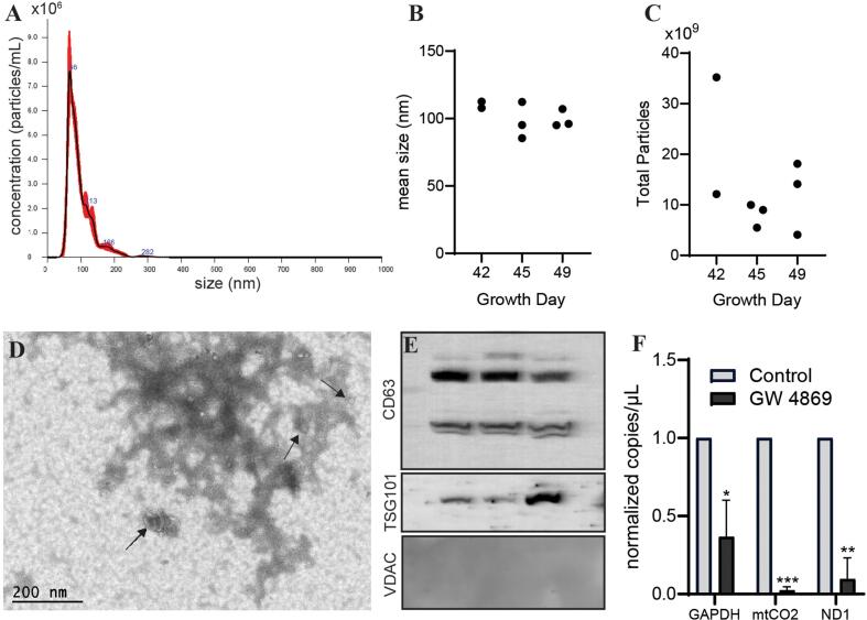
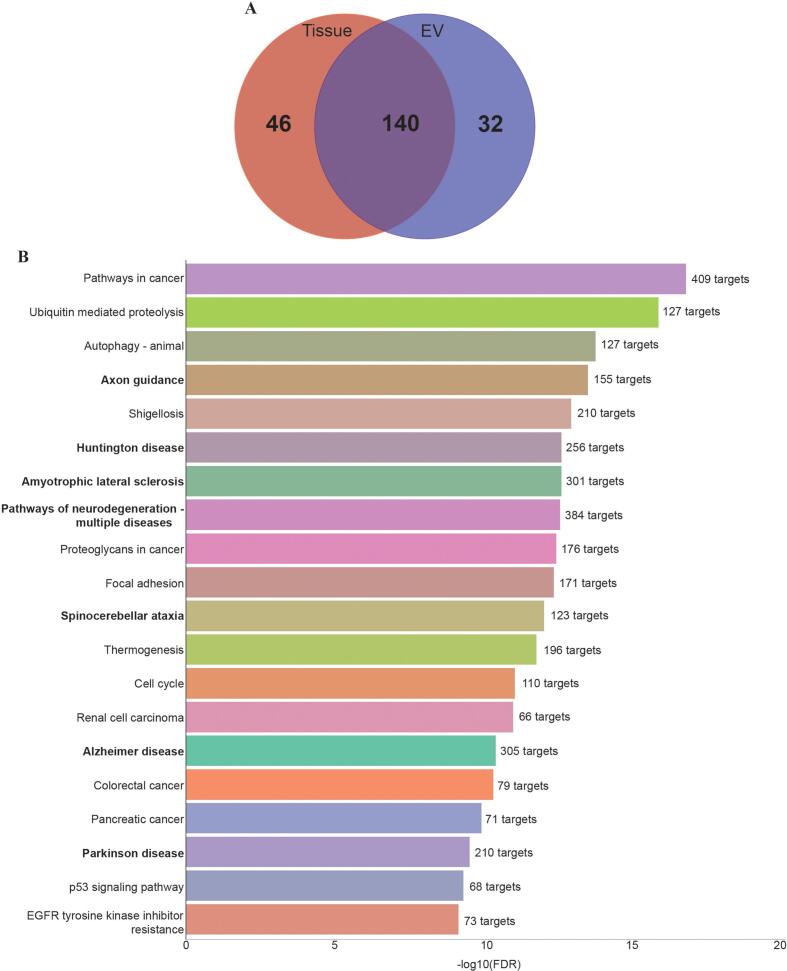
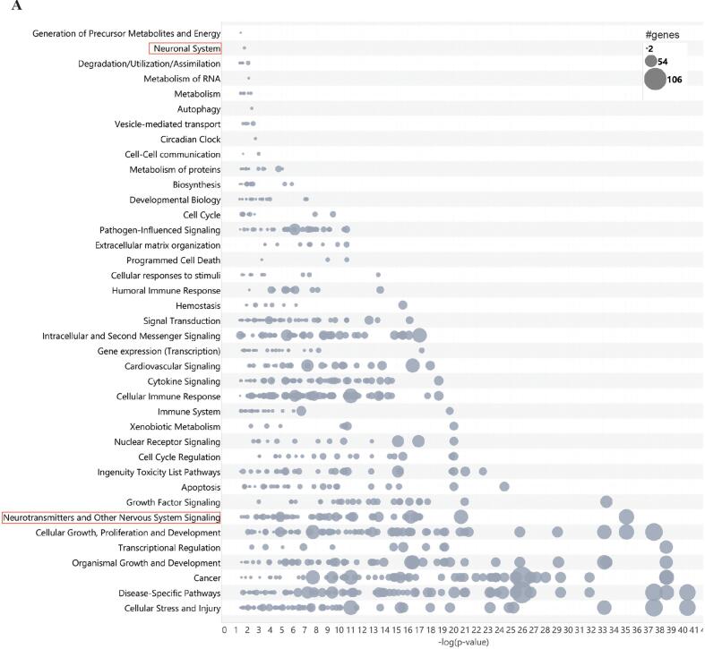
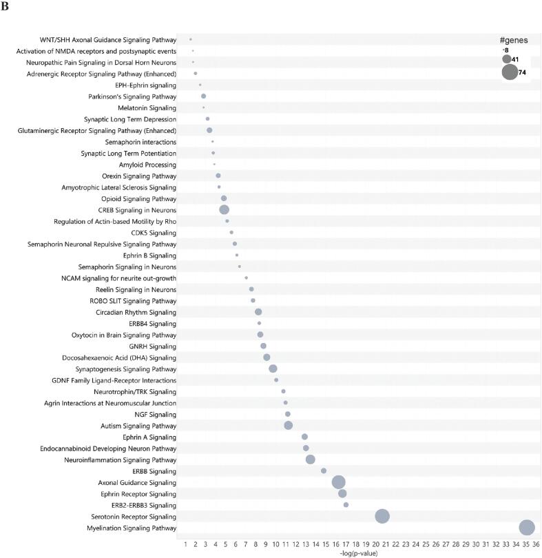
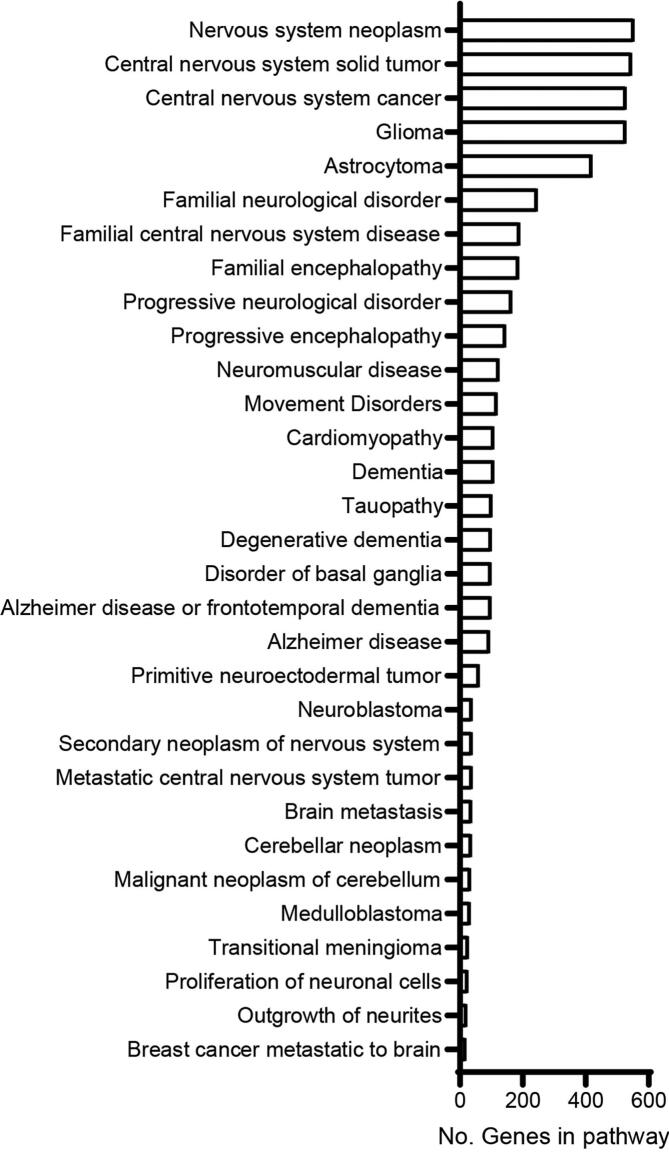
Environmental toxicants can contribute to the development of several neurodegenerative diseases. However, the mechanisms behind this pathology are still incompletely understood. Prompt diagnosis of impending neurodegeneration is crucial for early interventions to prevent cognitive decline. Towards this end, accurate biomarkers for early neurodegenerative processes and exposure risk are needed. Extracellular vesicles (EVs) are lipid particles released by cells which contain many bioactive molecules including miRNAs. EVs may serve both as a route of propagating neurotoxic phenotypes and as a source of biomarkers for neurological disease. However, the exact mechanisms though which EVs could spread the deleterious effects of toxicants and the full spectrum of their usage as biomarkers remain unclear. Organoid models have several advantages, including potential for use in high-throughput toxicant testing and applications in personalized medicine and disease models. However, few studies have examined EV release in brain organoids to determine if the EVs could contain useful biomarkers. We employed several technologies to characterize EVs released by human cerebral organoids and their associated miRNAs. We identified that cerebral organoids consistently release EV-associated miRNA in quantities sufficient for robust analysis with NanoString. Further, pathway analyses revealed that terms related to neurodegenerative disease and nervous system signaling are associated with the recovered miRNAs. Together, these data suggest that cerebral organoids have utility as a tool for the discovery of EV-associated miRNAs involved in neurodegenerative disease and neurotoxicity.
Keywords: Cerebral organoids; Extracellular vesicles; Neurodegenerative disease; Neurotoxicity; New approach methodologies; microRNA.
Conflict of interest statement
The authors declare that they have no known competing financial interests or personal relationships that could have appeared to influence the work reported in this paper.
Figures
Similar articles
-
SIV Infection Regulates Compartmentalization of Circulating Blood Plasma miRNAs within Extracellular Vesicles (EVs) and Extracellular Condensates (ECs) and Decreases EV-Associated miRNA-128.Viruses. 2023 Feb 24;15(3):622. doi: 10.3390/v15030622. Viruses. 2023. PMID: 36992331 Free PMC article.
-
Alterations in Abundance and Compartmentalization of miRNAs in Blood Plasma Extracellular Vesicles and Extracellular Condensates during HIV/SIV Infection and Its Modulation by Antiretroviral Therapy (ART) and Delta-9-Tetrahydrocannabinol (Δ9-THC).Viruses. 2023 Feb 24;15(3):623. doi: 10.3390/v15030623. Viruses. 2023. PMID: 36992332 Free PMC article.
-
EV-miRNAs from breast cancer patients of plasma as potential prognostic biomarkers of disease recurrence.Heliyon. 2024 Jul 10;10(14):e33933. doi: 10.1016/j.heliyon.2024.e33933. eCollection 2024 Jul 30. Heliyon. 2024. PMID: 39104474 Free PMC article.
-
Plasma neuronal exosomes serve as biomarkers of cognitive impairment in HIV infection and Alzheimer's disease.J Neurovirol. 2019 Oct;25(5):702-709. doi: 10.1007/s13365-018-0695-4. Epub 2019 Jan 4. J Neurovirol. 2019. PMID: 30610738 Free PMC article. Review.
-
Harnessing microRNA-enriched extracellular vesicles for liquid biopsy.Front Mol Biosci. 2024 Feb 21;11:1356780. doi: 10.3389/fmolb.2024.1356780. eCollection 2024. Front Mol Biosci. 2024. PMID: 38449696 Free PMC article. Review.
References
-
- Aharon A., Spector P., Ahmad R.S., Horrany N., Sabbach A., Brenner B., Aharon-Peretz J. Extracellular vesicles of Alzheimer's disease patients as a biomarker for disease progression. Mol. Neurobiol. 2020;57(10):4156–4169. - PubMed
-
- Arrifano G.D., Augusto-Oliveira M., Sealey-Bright M., Zainal J., Imbiriba L., Fernandes L.M.P., Maia C.S.F., Anthony D., Crespo-Lopez M.E. Contributing to understand the crosstalk between brain and periphery in methylmercury intoxication: neurotoxicity and extracellular vesicles. Int. J. Mol. Sci. 2021;22:19. - PMC - PubMed
LinkOut - more resources
Full Text Sources
