Case Reports
. 2025 May 5;17(5):e83488.
doi: 10.7759/cureus.83488. eCollection 2025 May.

Sclerosing Angiomatoid Nodular Transformation of the Spleen: Multimodality Imaging Features

Affiliations
Case Reports

Sclerosing Angiomatoid Nodular Transformation of the Spleen: Multimodality Imaging Features

Burhan Vural et al. Cureus. .

Abstract

Sclerosing angiomatoid nodular transformation (SANT) of the spleen is a rare benign lesion. This report presents the case of a middle-aged woman with asymptomatic SANT of the spleen, complicated by metastatic papillary thyroid carcinoma and an undiagnosed splenic mass. The condition was effectively managed through laparoscopic splenectomy, with a definitive diagnosis confirmed postoperatively. This case aims to contribute to enhancing the differential diagnosis of SANT by highlighting its radiological features, particularly in patients undergoing follow-up for malignancy.

Keywords: computed tomography (ct) imaging; magnetic resonance imaging(mri); sclerosing angiomatoid nodular transformation; spleen; ultrasound imaging.


Conflict of interest statement

Human subjects: All authors have confirmed that this study did not involve human participants or tissue. Conflicts of interest: In compliance with the ICMJE uniform disclosure form, all authors declare the following: Payment/services info: All authors have declared that no financial support was received from any organization for the submitted work. Financial relationships: All authors have declared that they have no financial relationships at present or within the previous three years with any organizations that might have an interest in the submitted work. Other relationships: All authors have declared that there are no other relationships or activities that could appear to have influenced the submitted work.

Figures

Figure 1
Figure 1. Gray-scale axial abdominal ultrasonography examination
At the level of the splenic hilum, a nodular lesion measuring approximately 26x29 mm with mildly lobulated contours is observed. It appears iso-hypoechoic compared to the surrounding splenic parenchyma (red arrows).
Figure 2
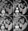
Figure 2. Dynamic contrast-enhanced axial CT examination
On non-contrast axial CT imaging (a), a hypodense mass lesion measuring 26x20 mm with mildly lobulated contours is observed in the inferior spleen (red arrows). On post-contrast images (red arrows), the lesion demonstrates peripheral nodular enhancement during the arterial phase (b) (red asterisks) and progressive centripetal enhancement during the portal phase (c). In the late venous phase images (d), the lesion shows enhancement similar to the surrounding splenic parenchyma.
Figure 3
Figure 3. Dynamic MRI examination
On non-contrast fat-suppressed T1-weighted axial imaging (a) and T2-weighted axial imaging (b), the lesion (black arrows) demonstrates iso-hypointense signal characteristics. On diffusion-weighted imaging (c) and ADC imaging (d), no significant diffusion restriction is observed. In T2-weighted imaging (b) and diffusion-weighted imaging (c), a centrally located hypointense scar (red asterisks) is noted. Following the administration of contrast material, on arterial T1-weighted imaging (e), the lesion (yellow arrows) demonstrates weak peripheral enhancement (red asterisks) compared to the splenic parenchyma. On post-contrast portal venous phase (f), delayed venous phase axial (g), and coronal (h) images, the lesion exhibits progressive centripetal enhancement (yellow arrows). In delayed venous phase images (g, h), hypointense signal characteristics consistent with a central scar are observed (black arrows).

References

    1. Sclerosing angiomatoid nodular transformation (SANT) of the spleen: Case report and review of the literature. Falk GA, Nooli NP, Morris-Stiff G, et al. Int J Surg Case Rep. 2012;3:492–500. - PMC - PubMed
    1. Sclerosing angiomatoid nodular transformation (SANT): report of 25 cases of a distinctive benign splenic lesion. Martel M, Cheuk W, Lombardi L, et al. Am J Surg Pathol. 2004;28:1268–1279. - PubMed
    1. Sclerosing angiomatoid nodular transformation of the spleen: lessons from a rare case and review of the literature. Nomura R, Tokumura H, Katayose Y, et al. Intern Med. 2019;58:1433–1441. - PMC - PubMed
    1. Sclerosing angiomatoid nodular transformation of the spleen: a report of rare case and literature review. Raja F, Kumar V, Moll E, et al. Cureus. 2023;15:0. - PMC - PubMed
    1. Solid tumors of the spleen: evaluation and management. Silver DS, Pointer DT Jr, Slakey DP. J Am Coll Surg. 2017;224:1104–1111. - PubMed

Publication types

LinkOut - more resources