A Radiographic Investigation Exploring Differences in Static Anterior Tibial Translation Expressed as a Percentage Between ACL-Deficient and -Intact Patients to Improve Interobserver Variability Between Different Centers
- PMID: 40470516
- PMCID: PMC12130644
- DOI: 10.1177/23259671251330310
A Radiographic Investigation Exploring Differences in Static Anterior Tibial Translation Expressed as a Percentage Between ACL-Deficient and -Intact Patients to Improve Interobserver Variability Between Different Centers
Abstract
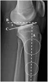
Background: Static anterior tibial translation (SATT) represents the amount of anterior translation due to axial load. It has been shown to be increased with anterior cruciate ligament (ACL) rupture, meniscal tear, and increased posterior tibial slope (PTS). It has also been shown to be correlated with ACL reconstruction failure. ACL reconstruction alone does not improve SATT. A sagittal plane slope-correcting osteotomy improves SATT, and SATT has recently been used to define the target slope correction after osteotomy. However, absolute values for SATT differ between institutions by >5 mm. Absolute measures differ based on the amount of magnification of the image, which varies based on the radiographic source to image distance, the source to object distance, rotation, and whether the medial or lateral condyle is presented to the source first. Scaled, or percentage radiographic measures, should correct for these differences.
Purpose: To express SATT as a percentage (SATT%) of the medial plateau distance to improve accuracy and interinstitutional utilization of SATT.
Study design: Cross-sectional study; Level of evidence, 3.
Methods: A consecutive series of patients without ligamentous or meniscal injuries between 2019 and 2022 was reviewed. A matched consecutive cohort of patients with nonacute ACL injuries (surgery between 6 and 12 weeks after injury) without concomitant pathology was reviewed. Preoperative SATT and PTS were measured with a previously validated technique on lateral weightbearing knee radiographs. Regression analysis was performed to investigate the relationship between SATT% and PTS.
Results: There were 101 controls and 115 patients with an ACL injury who were included in this study. In the control cohort, the mean SATT% was 3.18% (SD, 5.92%) and mean PTS was 10.61° (SD, 3.28°). This was significantly different from our ACL cohort's mean SATT% of 5.16% (SD, 7.41%) (P = .04) and mean PTS of 9.46° (2.85°) (P = .02). Linear regression analysis showed that for every 1° increase in PTS, there was a 0.08% increase in SATT% in the control cohort, so every 10° rise in slope was associated with an 0.8% increase in SATT%. In the ACL cohort, the effect of PTS on SATT% was larger; for every 1° of increase in PTS, there was an increase of 0.97% SATT%.
Conclusion: The present study reports a reference SATT% value of 3.18% (SD, 5.92%) in a non-ACL injured cohort, which was lower than the ACL cohort's mean 5.16% (SD 7.41%), despite the ACL cohort's having a longer medial tibial plateau than the control population. The effect of slope on weightbearing anterior tibial translation was greater in the ACL population compared with the control cohort. These scaled, percentage values should improve the interinstitutional usage of SATT.
Keywords: anterior cruciate ligament reconstruction; deflexion tibial osteotomy; radiograph; static anterior tibial translation; tibial slope.
© The Author(s) 2025.
Conflict of interest statement
The authors declared that there are no conflicts of interest in the authorship and publication of this contribution. AOSSM checks author disclosures against the Open Payments Database (OPD). AOSSM has not conducted an independent investigation on the OPD and disclaims any liability or responsibility relating thereto. Ethical approval for this study was obtained from GCS Ramsay Sante pour l’Enseignement et la Recherche (COS-RGDS-2022-09-008-DEMEY-G).
Figures


References
-
- Alqahtani Y, Murgier J, Beaufils P, Boisrenoult P, Steltzlen C, Pujol N. Anterior tibial laxity using the GNRB device in healthy knees. Knee. 2018;25(1):34-39. - PubMed
-
- Anderson AF, Irrgang JJ, Kocher MS, Mann BJ, Harrast JJ; International Knee Documentation Committee. The International Knee Documentation Committee Subjective Knee Evaluation Form: normative data. Am J Sports Med. 2006;34(1):128-135. - PubMed
-
- Cance N, Dan MJ, Pineda T, Demey G, Dejour DH. Radiographic investigation of differences in static anterior tibial translation with axial load between isolated ACL injury and controls. Am J Sports Med. 2024;52(2):338-343. - PubMed
-
- Dan MJ, Cance N, Pineda T, Demey G, Dejour DH. Four to 6 degrees is the target posterior tibial slope after tibial deflection osteotomy according to the knee static anterior tibial translation. Arthroscopy. 2024;40(3):846-854. - PubMed
-
- Dejour D, La Barbera G, Pasqualotto S, et al. Sagittal plane corrections around the knee. J Knee Surg. 2017;30(8):736-745. - PubMed
LinkOut - more resources
Full Text Sources

