. 2025 Dec;17(1):2515480.
doi: 10.1080/19490976.2025.2515480. Epub 2025 Jun 5.

L-serine promotes pro-carcinogenic effects of colibactin-producing E. coli

Affiliations

L-serine promotes pro-carcinogenic effects of colibactin-producing E. coli

Amandine Devaux et al. Gut Microbes. 2025 Dec.

Abstract

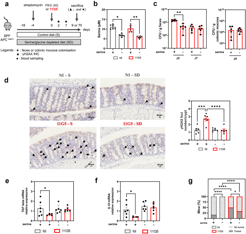
Colonic tissues are abnormally colonized by colibactin-producing Escherichia coli (CoPEC) in colorectal cancer (CRC) patients. CoPECs have been shown to promote colorectal carcinogenesis in several pre-clinical CRC mouse models. Here, we report that CoPEC reprograms the metabolism of colonic epithelial cells in a colibactin-dependent manner, leading to a Warburg-like effect, altered redox homeostasis, and disrupted amino acid metabolism. Among these metabolic modifications, we observed a significant decrease in both extracellular and intracellular serine levels. We found that CoPEC activates the L-serine-utilization operon during gut colonization, maximizing its competitive fitness advantage over a commensal strain. Moreover, an L-serine-depleted diet induces an early and transient decrease in CoPEC colonization of mice gut, associated with decrease of both DNA damages and tumor development. Finally, deletion of the bacterial tdcA gene involved in L-serine operon utilization reduces the competitive fitness of CoPEC, the in vitro adhesion and persistence within the epithelial cells and leads in CRC animal models to reduced carcinogenic activity of the pathobiont. This work highlights the interplay between intestinal microbiota factors, such as CoPEC, and nutritional factors, such as L-serine, in colorectal carcinogenesis.

Keywords: CoPEC; Colorectal cancer; L-serine; colibactin producing E. coli; pks island.


Conflict of interest statement

No potential conflict of interest was reported by the author(s).

Figures

None
Graphical abstract
Figure 1.
Figure 1.
Untargeted metabolomic profiling of T84 intracellular metabolites revealed a significant metabolome reprogramming following CoPEC infection in a colibactin-dependent manner. (a) Metabolomic experimental workflow. Non-targeted profiling of the intracellular metabolome from non-infected T84 cells and cells infected with E. coli 11G5 or 11G5-ΔclbQ strains (MOI 100) at 24 h post-infection (n = 10 per group). (b–d) Principal component analysis score plot from metabolomic data obtained by (b) 1H NMR, (c) LC-MS in positive and (d) negative modes. (e) Two-way hierarchical clustering heatmap of the 22 identified intracellular metabolites selected from the three PLS-DA model VIP score with a threshold of 1 between the three group comparisons. Branch lengths in dendrograms produced from cluster analysis correspond to the relative degree of similarity between branches. Differential variable intensity is represented for all variables as a color gradient across all samples from blue (lowest) to red (highest). (f) Metabolic set enrichment analysis of the 22 identified intracellular metabolites selected from the three PLS-DA model with a VIP score > 1 between the three group comparisons. (g) Box-plots based on intracellular discriminant metabolites selected from PLS-DA model with a VIP score with a threshold of 1 and with a p-value <0.05 between NI and 11G5 conditions and between 11G5 and 11G5-ΔclbQ conditions showing the pathways influenced by 11G5 infection. *p < 0.05; **p < 0.01; ***p < 0.001; ****p < 0.0001.
Figure 2.
Figure 2.
Activation of the L-serine-utilization-genes in CoPEC provided a growth advantage over a commensal strain during cell infection. (a–f) Analysis of the expression of genes involved in L-serine utilization or synthesis between (a, c, e) 11G5 CoPEC strain and K12 E. coli commensal strain and between (b, d, f) 11G5 and 11G5-ΔclbQ CoPEC strain 4 h after infection in T84 cells (MOI 100). mRNA expression of (a,b) serA gene coding for the synthesis of L-serine in E. coli. mRNA expression of two genes (c,d) tdcA and (e,f) tdcR coding for activators of the tdc operon involved in serine-utilization in E. coli. These data were normalized by the bacterial tufA translation elongation gene. (g,h) Growth curve (g) and its AUC (h) of 11G5 and K12 strains grown at 37°C without shaking in minimal medium (MM) containing 0.1% glucose and thiamine (1 mg/mL) and supplemented or not with L-serine (10 mm - sigma S4500 – SLCG1409). Bacterial growth was quantified by measuring optical density (O.D.600) every hour for 10 hours using the Flexstation 3 (multi-mode microplate Reader). Data are from a representative experiment performed at least three times and values represent mean ± standard error of the mean. *p < 0.05; **p < 0.01; ****p < 0.0001.
Figure 3.
Figure 3.
L-Serine depleted diet-fed induced a diminution of CoPEC genotoxic and pro-carcinogenic effects in the colon of APCmin/+ mice. (a) Experimental workflow of treatment and infection used in an SPF APCmin/+ mice model for this study: 6 mice per group. Mice were killed 9 d postinfection (p.i.) for short-time experiments (b–f) and 70 d p.i. for long-time experiments (g). (b) Serum serine concentration determined by liquid chromatography-mass spectrometry (LC-MS/MS) technique from a blood sample taken from the retro-orbital sinus during sacrifice. Each point represents the serine concentration of one mouse. (c) Level of colonization measured in the stools 2- and 7-d p.i. and in colonic mucosa sample taken at sacrifice at 9-d p.i. of mice infected with the 11G5 CoPEC strain. Results are presented in CFU/g or “colony-forming unit” for each condition. (d) γH2AX immunohistochemical staining of colonic mucosa and quantification of γH2AX foci number/crypt determined from 30 crypts/mouse. (e,f) mRNA expression of two genes coding for anti-inflammatory cytokines TGF beta (f) and -IL10 (f) in colon of mice. These data were normalized by the mouse S26 ribosomal protein gene. (g) Percentage of APCmin/+ mice developing colonic polyps 70 d p.i. Data are from a representative experiment performed at least two times. Values represent mean ± standard error of the mean or median ±95% of CI. *p < 0.05; **p < 0.01; ***p < 0.001; ****p < 0.0001.
Figure 4.
Figure 4.
The tdc operon provided a selective advantage to CoPEC. (a) Serum serine concentration determined by liquid chromatography-mass spectrometry (LC-MS/MS) technique in the intracellular medium of non-infected and infected T84 epithelial cells infected with 11G5 CoPEC strain and its corresponding mutant unable to metabolize L-serine 11G5-ΔtdcA. (b) Adhesion (3 h) internalization (4 h), survival (24 h) and persistence (48 h) assay of 11G5 CoPEC strain or 11G5-ΔtdcA in T84 cells (multiplicity of infection 100). Results are presented in CFU/mL or “colony-forming unit” for each condition. (c) Percentage of survival and bacterial persistence in T84 cells. (d) Agglutination test of 11G5 and its 11G5-ΔclbQ or 11G5-ΔtdcA mutants. LF82 and LF82-ΔfimH were used as positive and negative controls respectively. (e) Co-infection of 11G5 CoPEC strain and its mutant11G5-ΔtdcA in T84 cells at 3 h, 4 h, 24 h and 48 h post-infection (multiplicity of infection 10). (f) Experimental workflow of treatment and co-infection in SPF WT (C57Bl/6J) mice and SPF APCmin/+ (C57Bl/6J) mice model used in this study: between eight and nine mice per group. (g, h) Level of colonization measured in the stools and in the mucosa colonic in WT (g) and APCmin/+ (h) mice co-infected with the 11G5 and 11G5-ΔtdcA during experiment. Data are from a representative experiment performed at least three times for in vitro model and at least two times for in vivo model. Values represent mean ± standard error of the mean or median ±95% of CI. *p < 0.05; **p < 0.01; ***p < 0.001.
Figure 5.
Figure 5.
Utilization of L-serine by CoPEC via tdc operon promoted persistence, genotoxic effects and tumoral development in MC38 grafted model and in APCmin/+ model. (a) Experimental workflow of treatment and infection of SPF WT mice (C57Bl/6J) used in this study: between six and seven mice per group. (b) MC38 tumor growth in mice. (c) MC38 tumor volume at 12 d post-injection. (d) Experimental workflow of treatment and infection in SPF APCmin/+ mice model used in this study: between four or four mice per group. (e) Level of colonization and corresponding AUC measured in the stools of APCmin/+ mice infected with the 11G5 CoPEC strain or the corresponding mutant 11G5-ΔtdcA unable to metabolize L-serine during experiment. (f) Level of colonization measured in the mucosa colonic infected with the 11G5 or 11G5-ΔtdcA strain. (g) γH2AX immunohistochemical staining of colonic mucosa and quantification of γH2AX foci number/crypt determined from 30 crypts/mouse and four or five mice/group. (h) Number of visible colonic polyps and total size of polyps per mouse in infected mice with 11G5 or 11G5-ΔtdcA. Data are from a representative experiment performed at least two times. Values represent mean ± standard error of the mean or median ±95% of CI. *p < 0.05; ****p < 0.0001.

References

    1. Cancer (IARC) TIA for R on. Global Cancer Observatory [Internet]. [cited 2024 Jan 11]. https://gco.iarc.fr/.
    1. Wong CC, Yu J.. Gut microbiota in colorectal cancer development and therapy. Nat Rev Clin Oncol. 2023;20(7):429–17. doi: 10.1038/s41571-023-00766-x. - DOI - PubMed
    1. Dougherty MW, Jobin C. Intestinal bacteria and colorectal cancer: etiology and treatment. Gut Microbes. 2023;15(1):2185028. doi: 10.1080/19490976.2023.2185028. - DOI - PMC - PubMed
    1. Arthur JC, Perez-Chanona E, Mühlbauer M, Tomkovich S, Uronis JM, Fan T-J, Campbell BJ, Abujamel T, Dogan B, Rogers AB, et al. Intestinal inflammation targets cancer-inducing activity of the microbiota. Science. 2012;338(6103):120–123. doi: 10.1126/science.1224820. - DOI - PMC - PubMed
    1. Bonnet M, Buc E, Sauvanet P, Darcha C, Dubois D, Pereira B, Déchelotte P, Bonnet R, Pezet D, Darfeuille-Michaud A. Colonization of the human gut by E. coli and colorectal cancer risk. Clinical Cancer Research. 2014;20(4):859–867. doi: 10.1158/1078-0432.CCR-13-1343. - DOI - PubMed

MeSH terms