

Magnesium Resorbable Membrane for Guided Bone Regeneration in Critical Size Defect Model in Rabbits-Histomorphometric Analysis
- PMID: 40471118
- PMCID: PMC12139577
- DOI: 10.1111/cid.70055
Magnesium Resorbable Membrane for Guided Bone Regeneration in Critical Size Defect Model in Rabbits-Histomorphometric Analysis
Abstract
Objectives: To evaluate the effectiveness of a Mg-based membrane as a barrier for guided bone regeneration in rabbits calvaria.
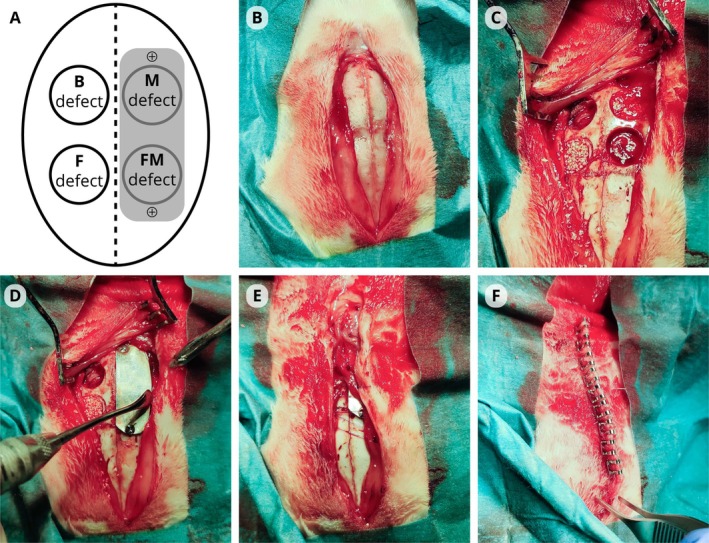
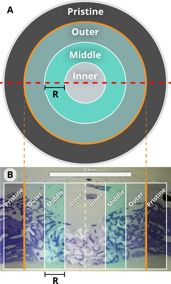
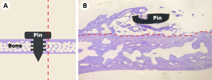
Materials and methods: Nine rabbits had four critical size defects created in each calvarium, randomly filled with a blood clot or bovine xenograft. One side was covered with the Mg-based membrane, and the control was left without membrane. After 8 weeks, histomorphometric analysis was performed to compare new bone formation with the pristine bone.
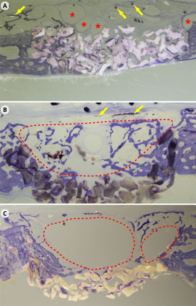
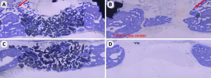
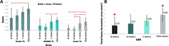
Results: Mg-supported membrane for guided bone regeneration (GBR) is safe and promotes bone formation in critical size defects (CSD) in a rabbit calvaria. Gas accumulation was observed in a third of the specimens due to membrane degradation. Histomorphometric analysis revealed greater bone formation in the defects grafted with the Mg membrane (4.85 ± 1.73 mm2) compared to the blood clot (2.14 ± 2.22 mm2). Treating lesions with filler or the magnesium membrane resulted in significantly higher bone formation in all three examined regions (25%-62%). New bone formation was observed beyond the original bone envelope.
Conclusion: Mg-based membrane supports guided bone regeneration (GBR), and when used in combination with a bone graft, enhances the performance despite the gas accumulation associated with membrane degradation.
Clinical relevance: Limited data exist on the use of Mg-based membranes in GBR and its effect on bone formation.
Keywords: critical size defects; guided bone regeneration; histomorphometry; magnesium; rabbits calvaria model; resorbable membrane.
© 2025 The Author(s). Clinical Implant Dentistry and Related Research published by Wiley Periodicals LLC.
Conflict of interest statement
The authors declare no conflicts of interest.
Figures
Similar articles
-
Guided bone regeneration in calvarial critical size bony defect using a double-layer resorbable collagen membrane covering a xenograft: a histological and histomorphometric study in rats.Oral Maxillofac Surg. 2018 Jun;22(2):203-213. doi: 10.1007/s10006-018-0694-x. Epub 2018 Apr 14. Oral Maxillofac Surg. 2018. PMID: 29654386
-
Bone healing with an in situ-formed bioresorbable polyethylene glycol hydrogel membrane in rabbit calvarial defects.Oral Surg Oral Med Oral Pathol Oral Radiol Endod. 2010 Mar;109(3):372-84. doi: 10.1016/j.tripleo.2009.10.008. Epub 2010 Jan 8. Oral Surg Oral Med Oral Pathol Oral Radiol Endod. 2010. PMID: 20060340
-
Guided bone regeneration using resorbable membrane and different bone substitutes: Early histological and molecular events.Acta Biomater. 2016 Jan;29:409-423. doi: 10.1016/j.actbio.2015.10.005. Epub 2015 Oct 13. Acta Biomater. 2016. PMID: 26441123
-
Research status and prospects of biodegradable magnesium-based metal guided bone regeneration membranes.Hua Xi Kou Qiang Yi Xue Za Zhi. 2024 Aug 1;42(4):415-425. doi: 10.7518/hxkq.2024.2024140. Hua Xi Kou Qiang Yi Xue Za Zhi. 2024. PMID: 39049628 Free PMC article. Review. Chinese, English.
-
Guided bone regeneration: materials and biological mechanisms revisited.Eur J Oral Sci. 2017 Oct;125(5):315-337. doi: 10.1111/eos.12364. Epub 2017 Aug 19. Eur J Oral Sci. 2017. PMID: 28833567 Free PMC article. Review.
References
-
- American Academy of Periodontology , Glossary of Periodontal Terms (American Academy of Periodontology, 1992).
-
- Wang H. L. and Boyapati L., ““PASS” Principles for Predictable Bone Regeneration,” Implant Dentistry 15, no. 1 (2006): 8–17. - PubMed
-
- Lang N. P., Berglundh T., Giannobile W. V., and Sanz M., Lindhe's Clinical Periodontology and Implant Dentistry (John Wiley & Sons, 2021).
-
- Witte F., “The History of Biodegradable Magnesium Implants: A Review,” Acta Biomaterialia 6, no. 5 (2010): 1680–1692. - PubMed
MeSH terms
Substances
Grants and funding
LinkOut - more resources
Full Text Sources
