

Bronchiectasis and airspace enlargement surrounding the lung nodule in dual-energy CT pulmonary angiography: comparison between iodine map and monochromatic image
- PMID: 40471409
- PMCID: PMC12339637
- DOI: 10.1007/s12194-025-00920-3
Bronchiectasis and airspace enlargement surrounding the lung nodule in dual-energy CT pulmonary angiography: comparison between iodine map and monochromatic image
Abstract
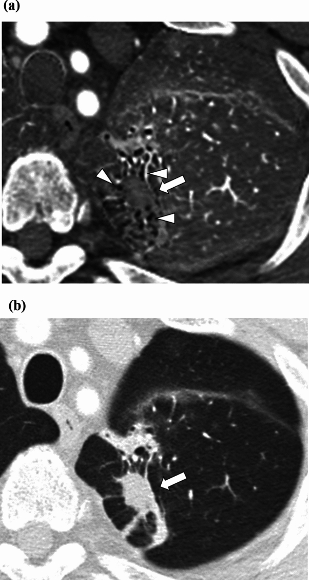
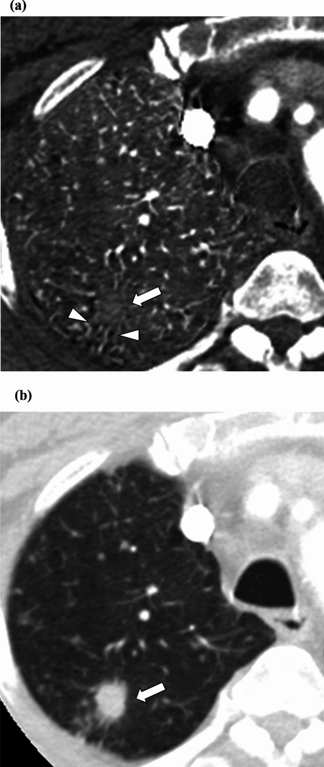
The purpose of the study is to investigate the degree and performance in the differential diagnosis of bronchiectasis/airspace enlargement in an iodine map obtainable from CT pulmonary angiography compared with monochromatic images. This retrospective study included 62 patients with a lung nodule who underwent CT pulmonary angiography. The iodine map and monochromatic image (70 keV) were reconstructed. Three readers evaluated the degree of bronchiectasis/airspace enlargement with a 4-point scale. A reference standard was established in 39 patients, and the performance of bronchiectasis/airspace enlargement in the differential diagnosis was evaluated in them. The degree of bronchiectasis/airspace enlargement in the iodine map (median score = 1/2/1 for reader 1/2/3) was significantly more prominent than that in the monochromatic image (median score = 0/1/0 for reader 1/2/3) (p < 0.001 for all readers). Using bronchiectasis/airspace enlargement, primary lung carcinoma and malignant lymphoma could be differentiated from other diseases, excluding lung infarct, with an area under the receiver operating characteristic curve (AUC) (reader 1/2/3) of 0.718/0.867/0.803 in the combinations of iodine map plus monochromatic image and 0.496/0.828/0.450 in the monochromatic image (p ≤ 0.047 for two readers). Lung metastasis from colorectal carcinoma could be differentiated from other diseases with an AUC of 0.851/0.976/0.838 in the combinations of iodine map plus monochromatic image, which was significantly superior to the monochromatic image (0.378/0.780/0.459) (p ≤ 0.012 for all readers). Bronchiectasis/airspace enlargement was more prominently observed in the iodine map than in the monochromatic image. This image finding in the iodine map provided added value in the differential diagnosis of malignant lung nodules compared with monochromatic images alone.
Keywords: Bronchiectasis; Lung cancer; Lung nodule; Multidetector computed tomography.
© 2025. The Author(s).
Conflict of interest statement
Declarations. Conflict of interest: The authors have no relevant financial or non-financial interests to disclose. Ethics approval: The Research Ethics Committee of the Faculty of Medicine of the University of Tokyo approved this retrospective study. This study adhered to the Declaration of Helsinki. Informed consent: The requirement for obtaining written informed consent was waived.
Figures


References
-
- Swensen SJ. CT screening for lung cancer. AJR Am J Roentgenol. 2002;179(4):833–6. 10.2214/ajr.179.4.1790833. - PubMed
-
- Thai AA, Solomon BJ, Sequist LV, Gainor JF, Heist RS. Lung cancer. Lancet. 2021;398(10299):535–54. 10.1016/S0140-6736(21)00312-3. - PubMed
-
- Herold CJ, Bankier AA, Fleischmann D. Lung metastases. Eur Radiol. 1996;6(5):596–606. 10.1007/BF00187656. - PubMed
Publication types
MeSH terms
Substances
LinkOut - more resources
Full Text Sources
