

Comparative clinical, virological and pathological characterization of equine rotavirus A G3P[12] and G14P[12] infection in neonatal mice
- PMID: 40471657
- PMCID: PMC12141749
- DOI: 10.1099/jgv.0.002110
Comparative clinical, virological and pathological characterization of equine rotavirus A G3P[12] and G14P[12] infection in neonatal mice
Abstract
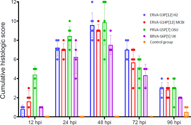
Group A rotavirus (RVA) infections are a leading cause of neonatal diarrhoea in foals. Neonatal mice could serve as a useful tool to study the pathogenesis of equine RVA (ERVA) as well as a preclinical model for assessment of vaccine efficacy. This study aimed to comparatively evaluate the clinical, virological and pathological features of ERVA G3P[12] and G14P[12] infection in neonatal mice and compare them with porcine OSU G5P[7] and bovine UK G6P[5] RVA reference strains. Neonatal mice orally inoculated with equine, bovine and porcine RVA developed short-lived diarrhoea at variable rates, G14P[12] (61%) and G3P[12] (88%). Viral replication kinetics for all strains were characterized by a gradual decline in viral load to levels below the limit of detection by 72-96 h post-infection (hpi), in line with the reduction in the number of infected enterocytes demonstrated via RNAscope® in situ hybridization. Importantly, the clinical and viral replication kinetics correlated with significant microscopic intestinal alterations characterized by enterocyte vacuolation, scalloping and hyperplasia with a peak occurring at 48 hpi and persisting until at least 96 hpi. Overall, neonatal mice develop a disease phenotype of short duration following infection with equine, porcine and bovine RVA strains characterized by diarrhoea and pronounced histological alterations in the intestinal villi. The limited intestinal viral replication is likely associated with host restriction. The clinical and pathological phenotypes developed by neonatal mice following experimental infection could serve as a preclinical tool to assess vaccine efficacy and for pathogenesis studies involving RVA of equine, porcine and bovine origin.
Keywords: G14P[12]; G3P[12]; bovine rotavirus A; equine rotavirus A; neonatal mice; porcine rotavirus A; rotavirus A; rotavirus tropism.
Conflict of interest statement
The authors declare that there are no conflicts of interest.
Figures


Similar articles
-
Genetic and antigenic characterization of two diarrhoeicdominant rotavirus A genotypes G3P[12] and G14P[12] circulating in the global equine population.J Gen Virol. 2024 Aug;105(8):002016. doi: 10.1099/jgv.0.002016. J Gen Virol. 2024. PMID: 39163114 Free PMC article.
-
Evaluation of inactivated vaccines against equine group A rotaviruses by use of a suckling mouse model.Vaccine. 2018 Sep 5;36(37):5551-5555. doi: 10.1016/j.vaccine.2018.07.057. Epub 2018 Jul 31. Vaccine. 2018. PMID: 30076106
-
Detection, molecular characterization and phylogenetic analysis of G3P[12] and G14P[12] equine rotavirus strains co-circulating in central Kentucky.Virus Res. 2018 Aug 15;255:39-54. doi: 10.1016/j.virusres.2018.05.025. Epub 2018 Jun 1. Virus Res. 2018. PMID: 29864502
-
Global distribution of group A rotavirus strains in horses: a systematic review.Vaccine. 2013 Nov 19;31(48):5627-33. doi: 10.1016/j.vaccine.2013.08.045. Epub 2013 Aug 28. Vaccine. 2013. PMID: 23994380
-
Equine rotavirus infection.J Equine Sci. 2021 Mar;32(1):1-9. doi: 10.1294/jes.32.1. Epub 2021 Mar 16. J Equine Sci. 2021. PMID: 33776534 Free PMC article. Review.
References
-
- Matthijnssens J, Ciarlet M, Heiman E, Arijs I, Delbeke T, et al. Full genome-based classification of rotaviruses reveals a common origin between human Wa-Like and porcine rotavirus strains and human DS-1-like and bovine rotavirus strains. J Virol. 2008;82:3204–3219. doi: 10.1128/JVI.02257-07. - DOI - PMC - PubMed
Publication types
MeSH terms
LinkOut - more resources
Full Text Sources
Medical