. 2025 Jul;12(4):e200414.
doi: 10.1212/NXI.0000000000200414. Epub 2025 Jun 5.

Glymphatic System May Mediate the Relation Between Choroid Plexus and Brain Damage in Multiple Sclerosis

Affiliations

Glymphatic System May Mediate the Relation Between Choroid Plexus and Brain Damage in Multiple Sclerosis

Paolo Preziosa et al. Neurol Neuroimmunol Neuroinflamm. 2025 Jul.

Abstract

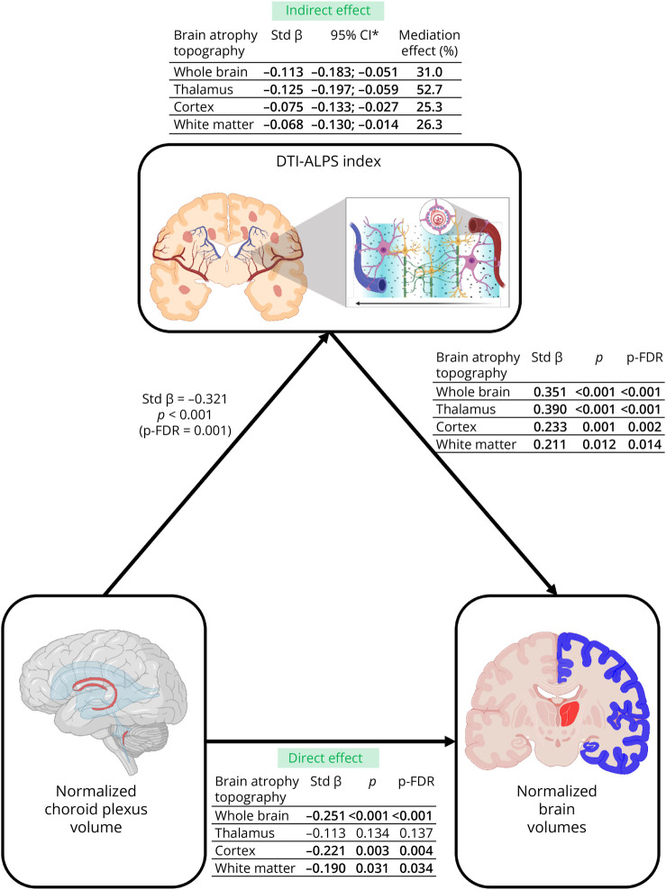
Background and objectives: The choroid plexus (CP) regulates immune functions and produces most CSF that circulates in the brain parenchyma through perivascular spaces, part of the glymphatic system. In multiple sclerosis (MS), CP enlargement and glymphatic dysfunction are associated with inflammatory activity, clinical disability, and brain damage, but their interrelation is unclear. We investigated whether glymphatic system dysfunction mediates the association between CP enlargement and brain damage in patients with MS.

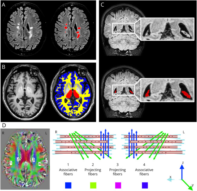
Methods: Brain fluid-attenuated inversion recovery, 3-dimensional T1-weighted, diffusion-weighted, and susceptibility-weighted sequences were obtained from 146 patients with MS and 72 healthy controls (HC). Glymphatic function was assessed using the diffusion along the perivascular space (DTI-ALPS) index, and CP volume was measured automatically.

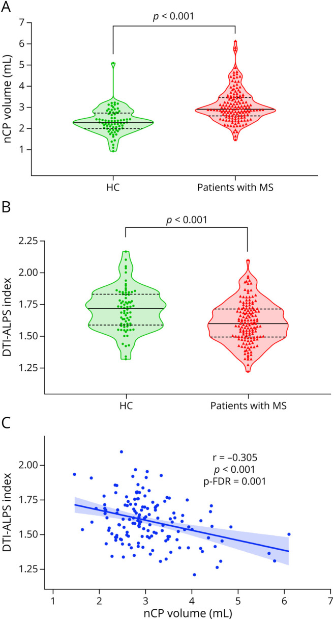
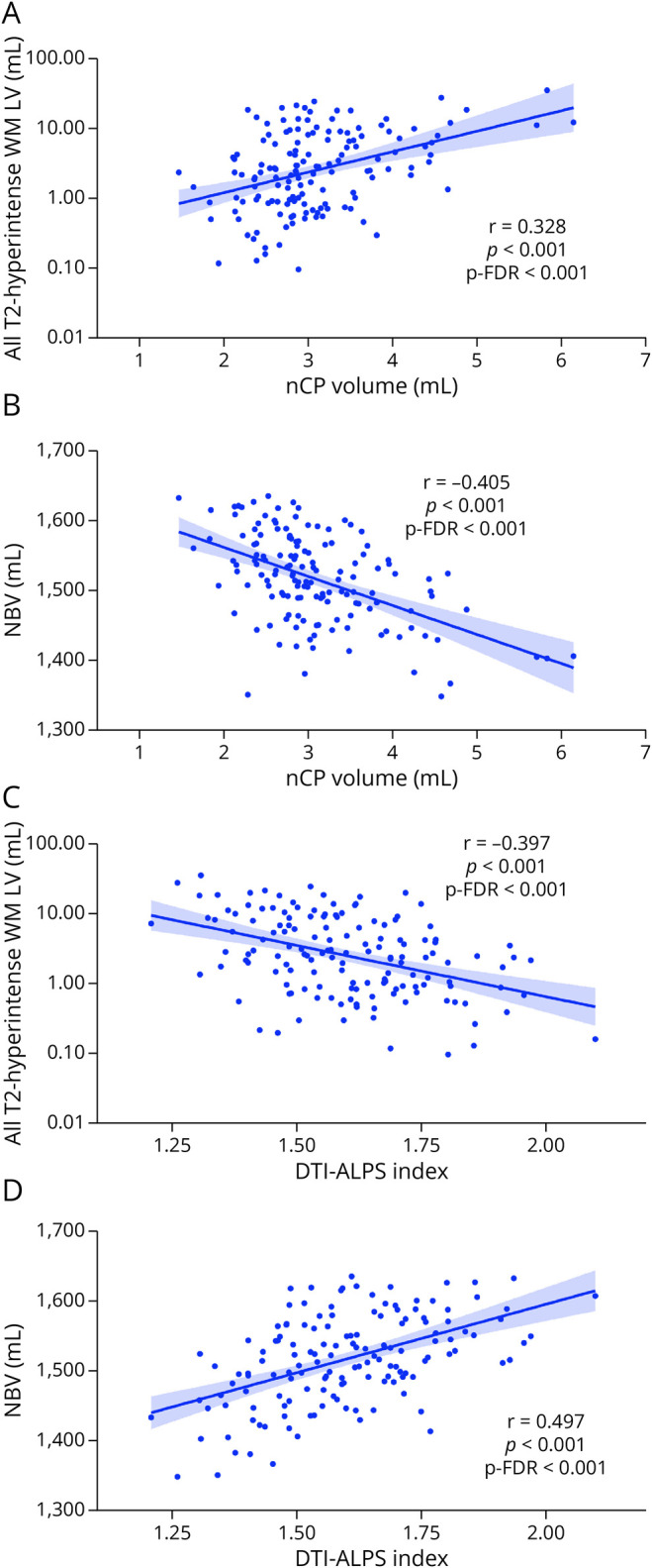
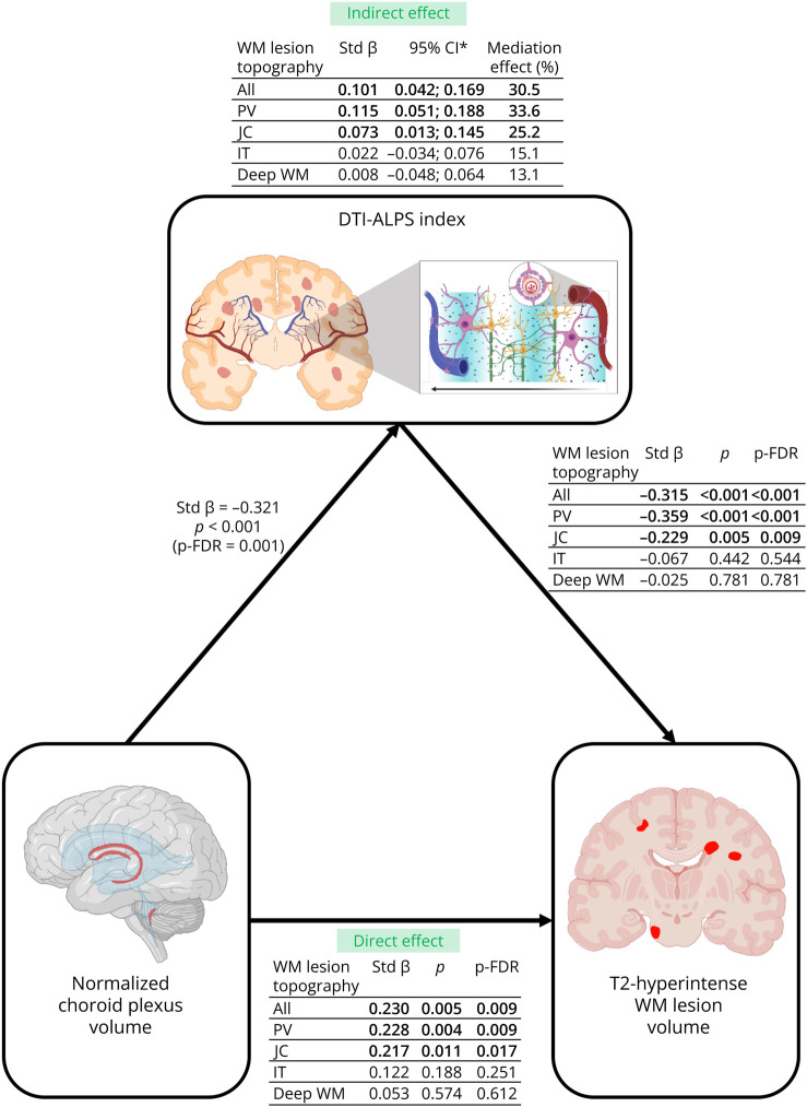
Results: Patients with MS showed significantly higher white matter (WM) lesion and CP volumes (p < 0.001), and lower DTI-ALPS index, brain, WM, thalamic, and cortical volumes than HC (p ≤ 0.048). In patients with MS, higher CP volume correlated with a lower DTI-ALPS index (r = -0.305, false discovery rate p value = 0.001). Both measures were associated with higher total, periventricular, and juxtacortical (JC) WM lesion volumes (CP volume: r from 0.285 to 0.340, p-FDR ≤ 0.001; DTI-ALPS index: r from -0.301 to -0.444, p ≤ 0.001), and lower brain, thalamic, cortical, and WM volumes (CP volume: r from -0.246 to -0.405, p-FDR ≤ 0.006; DTI-ALPS index: from 0.269 to 0.497, p-FDR ≤ 0.003). The DTI-ALPS index partially mediated the associations of normalized choroid plexus volume with total, periventricular, and JC T2-hyperintense WM lesion volumes (standardized-β ranging from 0.073 to 0.115, relative effect ranging from 25.2% to 33.6%) and normalized brain, thalamic, cortical, and WM volumes (standardized-β ranging from -0.086 to -0.125, relative effect ranging from 25.3% to 52.7%).

Discussion: In MS, enlarged normalized CP volume may contribute to brain damage accumulation possibly through the promotion of a chronic proinflammatory state and the mediation of glymphatic system dysfunction.


Conflict of interest statement

The authors declare that they have no competing interests in relation to this work. Potential conflicts of interest outside the submitted work are as follows: P. Preziosa received speaker honoraria from Roche, Biogen, Novartis, Merck, Bristol Myers Squibb, Genzyme, Horizon, and Sanofi; and has received research support from Italian Ministry of Health and Fondazione Italiana Sclerosi Multipla. E. Pagani has nothing to disclose. M. Margoni reports personal fees from Sanofi Genzyme, Merck Serono, Roche, Biogen, and Novartis. M. Rubin, L. Storelli, and G. Corazzolla have nothing to disclose. M.A. Rocca received consulting fees from Biogen, Bristol Myers Squibb, Eli Lilly, Janssen, and Roche; received speaker honoraria from AstraZaneca, Biogen, Bristol Myers Squibb, Bromatech, Celgene, Genzyme, Horizon Therapeutics Italy, Merck Serono SpA, Novartis, Roche, Sanofi, and Teva; receives research support from the MS Society of Canada, the Italian Ministry of Health, the Italian Ministry of University and Research, and Fondazione Italiana Sclerosi Multipla; and is an associate editor for Multiple Sclerosis and Related Disorders. M. Filippi is editor-in-chief of the Journal of Neurology, is an associate editor of Human Brain Mapping, Neurologic Sciences, and Radiology; received compensation for consulting services from Alexion, Almirall, Biogen, Merck, Novartis, Roche, and Sanofi; for speaking activities from Bayer, Biogen, Celgene, Chiesi Italia SpA, Eli Lilly, Genzyme, Janssen, Merck-Serono, Neopharmed Gentili, Novartis, Novo Nordisk, Roche, Sanofi, Takeda, and TEVA; for participation in Advisory Boards for Alexion, Biogen, Bristol-Myers Squibb, Merck, Novartis, Roche, Sanofi, Sanofi-Aventis, Sanofi-Genzyme, and Takeda; and for scientific direction of educational events for Biogen, Merck, Roche, Celgene, Bristol-Myers Squibb, Lilly, Novartis, and Sanofi-Genzyme; and receives research support from Biogen Idec, Merck-Serono, Novartis, Roche, the Italian Ministry of Health, the Italian Ministry of University and Research, and Fondazione Italiana Sclerosi Multipla. Full disclosure form information provided by the authors is available with the full text of this article at Neurology.org/NN.

Figures

Figure 1
Figure 1. Examples of WM Lesion, Brain Volumetric, and CP Segmentation and of DTI-ALPS Index Calculation
(A) Segmentation of brain T2-hyperintense WM lesions (red coded) on the 3D FLAIR sequence. (B) Segmentation of brain WM (yellow coded), cortex (blue coded), and thalamus (red coded) on 3D T1-weighted images using FSL-SIENAx software and FSL-FIRST tool. (C) CP segmentation (red coded) on the coronal plane of the 3D FLAIR sequence. (D) To calculate the DTI-ALPS index, the authors first registered the 3D FLAIR, the T2-hyperintense WM lesion mask and the FA map to the SWI scans. Three slices where veins run perpendicular to lateral ventricle were selected through SWI images. Over these slices, using the color-coded FA map in SWI space, four 3 × 3 × 3 mm regions of interest were drawn: 2 over the areas of associative fibers (see numbers 1 and 4), and the other 2 over the areas of projection fibers (see numbers 2 and 3) in the left and right hemispheres. Regions of interest were then registered to the diffusion space using the inverse of the FA map to the SWI registration matrix. Diffusivities along the x-, y-, and z-axes were extracted for each region of interest. DTI-ALPS was calculated as the ratio of diffusivities perpendicular to fiber bundles and parallel to veins (Dx proj and Dx assoc) over diffusivities perpendicular to fiber bundles and perpendicular to veins (Dy proj and Dz assoc). See text for further details. 3D = 3-dimensional; CP = choroid plexus; DTI-ALPS = diffusion tensor image analysis along the perivascular space; FA = fractional anisotropy; FLAIR = fluid attenuated inversion recovery; FSL = FMRIB Software Library; L = left; R = right; SWI = susceptibility-weighted imaging; WM = white matter.
Figure 2
Figure 2. Normalized Choroid Plexus Volume and DTI-ALPS Index Distributions in Patients With MS and HC and Partial Correlation Analyses Between Normalized Choroid Plexus Volume and DTI-ALPS Index in Patients With MS
Violin plots showing the distribution of (A) normalized choroid plexus volume and (B) DTI-ALPS index in patients with MS and HC. (C) Age-adjusted and sex-adjusted partial correlations between normalized choroid plexus volume and DTI-ALPS index in patients with MS. See text for further details. DTI-ALPS = diffusion tensor image analysis along the perivascular space; HC = healthy controls; mL = milliliter; MS = multiple sclerosis; nCP = normalized choroid plexus.
Figure 3
Figure 3. Partial Correlation Analyses in Patients With MS
Age-adjusted and sex-adjusted partial correlations in patients with MS between (A) normalized choroid plexus volume and all T2-hyperintense WM lesion volume, (B) normalized choroid plexus volume and NBV, (C) DTI-ALPS index and all T2-hyperintense WM lesion volume, and (D) DTI-ALPS index and NBV. See text for further details. DTI-ALPS = diffusion tensor image analysis along the perivascular space; mL = milliliter; LV = lesion volume; NBV = normalized brain volume; nCP = normalized choroid plexus; p-FDR = false discovery rate p value; WM = white matter.
Figure 4
Figure 4. Partial Mediation Effect of DTI-ALPS Index Between Choroid Plexus Volume and T2-Hyperintense WM Lesion Volumes in Patients With MS
The DTI-ALPS index partially mediated the association between choroid plexus volume and all, PV and JC WM lesion volumes. Mediation analysis was adjusted for age and sex. Created in BioRender. Preziosa, P. (2025)//BioRender.com/w88vafq. *Bias corrected and accelerated bootstrapped CI based on 5,000 samples. PROCESS (version 4.2) for SPSS primarily uses bootstrap CI to assess the significance of indirect effects instead of relying on traditional p values. If the 95% CI does not contain zero, the indirect effect is considered significant. Accordingly, all the direct effect with the 95% CI does not contain zero should be considered statistically significant (p < 0.05). DTI-ALPS = diffusion tensor image analysis along the perivascular space; IT = infratentorial; JC = juxtacortical; LV = lesion volume; MS = multiple sclerosis; p-FDR = false discovery rate p value; PV = periventricular; std β = standardized β; WM = white matter.
Figure 5
Figure 5. Partial Mediation Effect of DTI-ALPS Index Between Choroid Plexus Volume and Brain Volumes in Patients With MS
The DTI-ALPS index partially mediated the association between choroid plexus volume and normalized brain, thalamic, cortical, and WM volumes. Mediation analysis was adjusted for age and sex. Created in BioRender. Preziosa, P. (2025) BioRender.com/. *Bias corrected and accelerated bootstrapped CI based on 5,000 samples. PROCESS (version 4.2) for SPSS primarily uses bootstrap CI to assess the significance of indirect effects instead of relying on traditional p values. If the 95% CI does not contain zero, the indirect effect is considered significant. Accordingly, all the direct effect with the 95% CI does not contain zero should be considered statistically significant (p < 0.05). DTI-ALPS = diffusion tensor image analysis along the perivascular space; MS = multiple sclerosis; p-FDR = false discovery rate p value; std β = standardized β.

Similar articles

References

    1. Filippi M, Bar-Or A, Piehl F, et al. Multiple sclerosis. Nat Rev Dis Primers. 2018;4(1):43. doi: 10.1038/s41572-018-0041-4 - DOI - PubMed
    1. Lazarevic I, Soldati S, Mapunda JA, et al. The choroid plexus acts as an immune cell reservoir and brain entry site in experimental autoimmune encephalomyelitis. Fluids and Barriers of the CNS. 2023;20(1):39. doi: 10.1186/s12987-023-00441-4 - DOI - PMC - PubMed
    1. Fleischer V, Gonzalez-Escamilla G, Ciolac D, et al. Translational value of choroid plexus imaging for tracking neuroinflammation in mice and humans. Proc Natl Acad Sci U S A. 2021;118(36):e2025000118. doi: 10.1073/pnas.2025000118 - DOI - PMC - PubMed
    1. Bergsland N, Dwyer MG, Jakimovski D, et al. Association of choroid plexus inflammation on MRI with clinical disability progression over 5 Years in patients with multiple sclerosis. Neurology. 2023;100(9):e911-e920. doi: 10.1212/WNL.0000000000201608 - DOI - PMC - PubMed
    1. Ricigliano VAG, Morena E, Colombi A, et al. Choroid plexus enlargement in inflammatory multiple sclerosis: 3.0-T MRI and translocator protein PET evaluation. Radiology. 2021;301(1):166-177. doi: 10.1148/radiol.2021204426 - DOI - PubMed