. 2025 Sep 17;66(3):2301699.
doi: 10.1183/13993003.01699-2023. Print 2025 Sep.

Multi-omic spatial profiling reveals the unique SARS-CoV-2 lung microenvironment and collagen VI as a predictive biomarker in severe COVID-19

Affiliations

Multi-omic spatial profiling reveals the unique SARS-CoV-2 lung microenvironment and collagen VI as a predictive biomarker in severe COVID-19

Éanna Fennell et al. Eur Respir J. .

Abstract

Background: While coronavirus disease 2019 (COVID-19) is primarily a respiratory infection, few studies have characterised the immune response to COVID-19 in lung tissue. We sought to understand the pathogenic role of microenvironmental interactions and the extracellular matrix in post-mortem COVID-19 lung using an integrative multi-omic approach.

Methods: Post-mortem formalin-fixed paraffin-embedded lung tissue from fatal COVID-19 and nonrespiratory death control lung underwent multi-omic evaluation by Quantseq Bulk RNA sequencing, Nanostring GeoMx spatial transcriptomics, RNAscope, multiplex immunofluorescence and immunohistochemistry, to evaluate virus distribution, immune composition and the extracellular matrix. Markers of extracellular synthesis and breakdown were measured in the serum of 215 patients with COVID-19 and 54 healthy volunteer controls using ELISA.

Results: We found that severe acute respiratory syndrome coronavirus 2 (SARS-CoV-2) infection was restricted to the pneumocytes and macrophages of early-stage disease. Spatial analyses revealed an immunosuppressive virus microenvironment, enriched for PDL1+IDO1+ macrophages and depleted of T-cells. Oligoclonal T-cells in COVID-19 lung showed no enrichment of SARS-CoV-2 specific T-cell receptors. Collagen VI was upregulated and contributed to alveolar wall thickening and impaired gas exchange in COVID-19 lung. Serum from COVID-19 patients showed increased levels of PRO-C6, a marker of collagen VI synthesis, predicted mortality in hospitalised patients.

Conclusions: Our data refine the current model of respiratory COVID-19 with regard to virus distribution, immune niches and the role of the noncellular microenvironment in pathogenesis and risk stratification in COVID-19. We show that collagen deposition is an early event in the course of the disease.


Conflict of interest statement

Conflicts of interest: Serum ELISA assays were performed by Nordic Bioscience for no fee. J.M. Bülow Sand, M.A. Karsdal and D.J. Leeming are employees of Nordic Bioscience. Multiplex immunofluorescence (mIF) on the Akoya Phenocycler platform were performed by Akoya Biosciences for no fee. N. Nikulina, B.B. Cheikh and O. Braubach were employees of Akoya Biosciences. Initial analysis of the mIF was performed by A.T. Mayer of Enable Medicine for no fee. A.T. Mayer is president, chief scientific officer and founder of Enable Medicine. The remaining authors have no potential conflicts of interest to disclose.

Figures

None
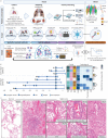
Overview of the study. SARS-CoV-2: severe acute respiratory syndrome coronavirus 2; COVID-19: coronavirus disease 2019; TCR: T-cell receptor.
FIGURE 1
FIGURE 1
Study design and temporal-clinicopathological features of tissue cohort. a) Schematic diagram of study workflow; b) disease timeline of coronavirus disease 2019 (COVID-19) cohort with associated clinical and pathological information; c) representative haematoxylin and eosin images of normal and COVID-19 lung tissue. MERS: Middle East respiratory syndrome; FFPE: formalin-fixed paraffin-embedded; TMA: tissue microarray; IHC: immunohistochemistry; mIF: multiplex immunofluorescence; ISH: in situ hybridisation; DSP: Digital Spatial Profiler; ITU: intensive therapy unit; BMI: body mass index; DAD: diffuse alveolar damage; OP: organising pneumonia; LV: lymphocytic vasculitis; SBI: secondary bacterial infection.
FIGURE 2
FIGURE 2
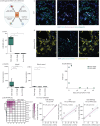
Severe acute respiratory syndrome coronavirus 2 (SARS-CoV-2) detection, distribution, features and validation. a) Schematic of multi-omic SARS-CoV-2 detection; b) quantification of SARS-CoV-2-infected cells; c) representative multiplex immunohistochemistry (mIHC) images of SARS-CoV-2 dual antibody detection; d) representative RNAscope fluorescence in situ hybridisation (FISH) images of SARS-CoV-2 infection in macrophages; e) localisation of SARS-CoV-2 infection predominantly to the alveoli by Nanostring GeoMx Digital Spatial Profiler (DSP); f) bulk RNA sequencing (RNAseq) viral load as a function of days post symptom onset of coronavirus disease 2019 (COVID-19); g) breakdown of viral gene expression across early- and late-stage COVID-19 by bulk RNAseq; h) cross-platform validation of SARS-CoV-2 detection (mIHC, RNAscope FISH and bulk RNAseq). mIF: multiplex immunofluorescence; DAPI: 4',6-diamidino-2-phenylindole. *: p<0.05; ****: p<0.0001.
FIGURE 3
FIGURE 3
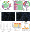
Immune landscape and T-cell repertoire of coronavirus disease 2019 (COVID-19) lung infection. a) Downsampled t-distributed stochastic neighbour embedding (tSNE) plot of identified cell types by multiplex immunohistochemistry (mIHC); b) cellular abundances of identified phenotypes per region analysed and pie chart of cell types present in COVID-19 lungs; c) representative images of the immune shift from early- to late-stage disease; d) Shannon diversity and richness of the COVID-19 tissue T-cell receptor (TCR) repertoire; e) clonal expansions of the tissue TCR repertoire; f) specificity of the tissue TCR repertoire. NK: natural killer; SMA: smooth muscle actin; Tregs: regulatory T-cells; APC: antigen-presenting cell; SARS-CoV-2: severe acute respiratory syndrome coronavirus 2; HLA: human leukocyte antigen; Mac: macrophage; Mono: monocyte; Epi; epithelial; MERS: Middle East respiratory syndrome; DAPI: 4',6-diamidino-2-phenylindole.
FIGURE 4
FIGURE 4
Immune and inflammatory virus microenvironment of severe acute respiratory syndrome coronavirus 2 (SARS-CoV-2). a) Immune cell abundance in the microenvironment of infected and control epithelium; b) immune cell abundance as a function of distance from SARS-CoV-2 infected epithelium and control epithelium; c) representative images and segmentation mask of SARS-CoV-2 microenvironment; d) differential gene expression of SARS-CoV-2-high versus -low alveoli by Nanostring GeoMx Digital Spatial Profiler (DSP); e) GO pathway analysis of SARS-CoV-2-positive alveoli; f) interleukin (IL)-6 expression by RNAscope in SARS-CoV-2-positive microenvironments; g) model of immune control and evasion in the SARS-CoV-2 microenvironment. mIF: multiplex immunofluorescence; HLA: human leukocyte antigen; DAPI: 4',6-diamidino-2-phenylindole; FISH: fluorescence in situ hybridisation; IL: interleukin; pDC: plasmacytoid dendritic cell.
FIGURE 5
FIGURE 5
Collagen landscape of fatal coronavirus disease 2019 (COVID-19). a) Lacunar space quantification; b) representative picosirius red images from early- and late-stage COVID-19; c) fibrotic predictive coefficient of genes; d) tissue-wide transcription of collagens in COVID-19 lungs and controls; e) tissue-wide transcription of type 1, 3, 4, 6 and 15 collagens in COVID-19 lungs on controls; f) spatially resolved collagen gene expression to alveoli and blood vessels; g) quantification of type 4 and 6 collagen deposition from multiplex immunohistochemistry (mIHC); h) representative images of type 6 collagen deposition in COVID-19 lungs by COMET mIHC. Scale bar=200 μm; i) correlation of collagen gene expression and macrophage activation. F: fibrosis; MERS: Middle East respiratory syndrome; DAPI: 4',6-diamidino-2-phenylindole. *: p<0.05; **: p<0.001; ***: p<0.001.
FIGURE 6
FIGURE 6
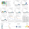
Blood detection of tissue-derived extracellular matrix landscape of coronavirus disease 2019 (COVID-19). a) Serum proteomics cohort description; b–h) collagen formation and degradation peptide serum levels for each group; i) survival cures for acute hospital patients based on serum collagen VI; j) survival cures for intensive therapy unit (ITU) patients based on serum collagen VI; k) demographic variable hazard ratios for outcome; l) blood-based biomarker of tissue derived collagen formation peptides for patient risk stratification. *: p<0.05; **: p<0.001; ***: p<0.001; ****: p<0.0001.

Comment in

References

    1. Watson OJ, Barnsley G, Toor J, et al. Global impact of the first year of COVID-19 vaccination: a mathematical modelling study. Lancet Infect Dis 2022; 22: 1293–1302. doi: 10.1016/S1473-3099(22)00320-6 - DOI - PMC - PubMed
    1. Herold T, Jurinovic V, Arnreich C, et al. Elevated levels of IL-6 and CRP predict the need for mechanical ventilation in COVID-19. J Allergy Clin Immunol 2020; 146: 128–136. doi: 10.1016/j.jaci.2020.05.008 - DOI - PMC - PubMed
    1. Liu Z, Li J, Chen D, et al. Dynamic interleukin-6 level changes as a prognostic indicator in patients with COVID-19. Front Pharmacol 2020; 11: 1093. doi: 10.3389/fphar.2020.01093 - DOI - PMC - PubMed
    1. Scherger S, Henao-Martínez A, Franco-Paredes C, et al. Rethinking interleukin-6 blockade for treatment of COVID-19. Med Hypotheses 2020; 144: 110053. doi: 10.1016/j.mehy.2020.110053 - DOI - PMC - PubMed
    1. Moss P. The T cell immune response against SARS-CoV-2. Nat Immunol 2022; 23: 186–193. doi: 10.1038/s41590-021-01122-w - DOI - PubMed