

Quantitative [89Zr]Zr-Trastuzumab PET and Diffusion- Weighted MRI for Characterization of Metastatic HER2-Positive Breast Cancer with PET/MRI
- PMID: 40473459
- PMCID: PMC12212413
- DOI: 10.2967/jnumed.124.268931
Quantitative [89Zr]Zr-Trastuzumab PET and Diffusion- Weighted MRI for Characterization of Metastatic HER2-Positive Breast Cancer with PET/MRI
Abstract
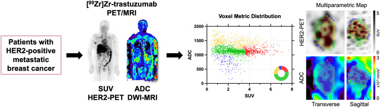
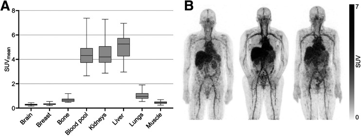
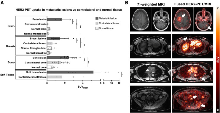
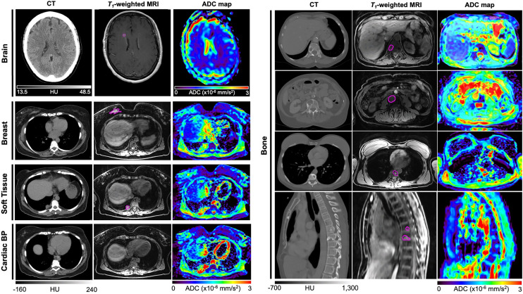
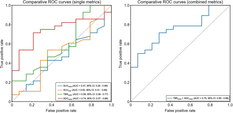
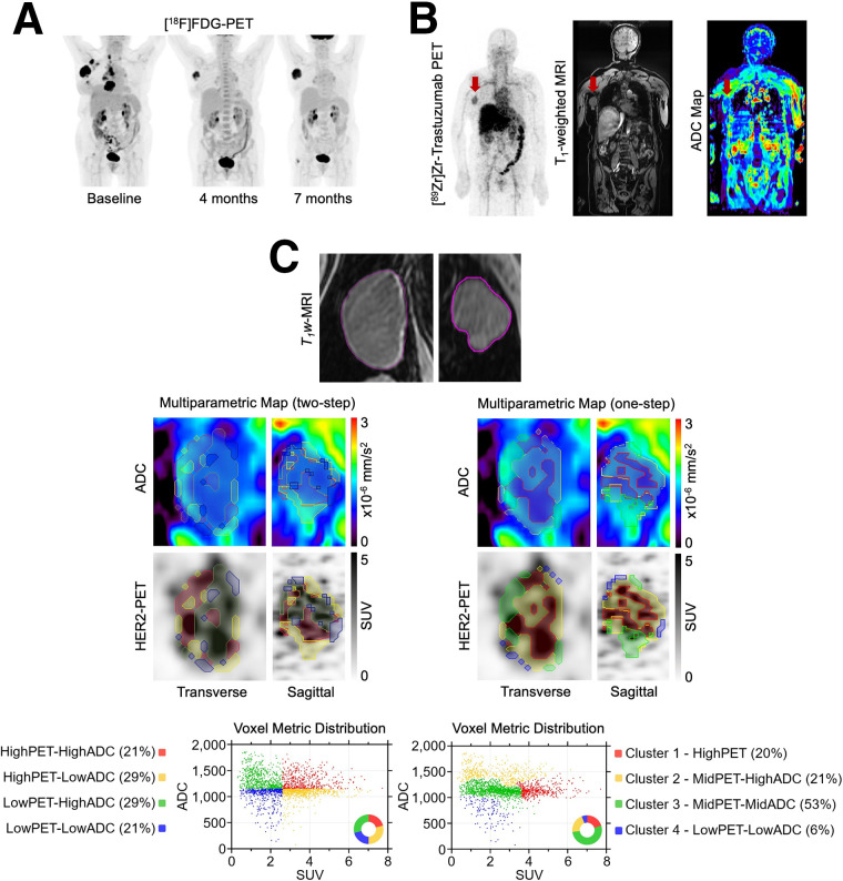
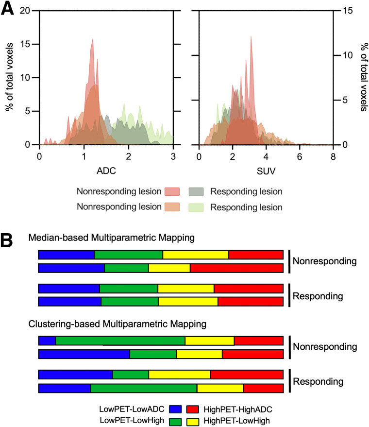
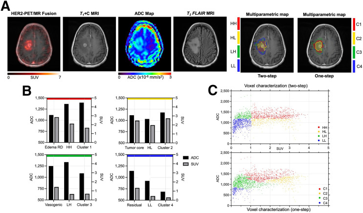
Current methods for evaluating HER2 expression in breast cancer are invasive and fail to capture spatial and temporal heterogeneity between primary tumors and metastases. Nuclear imaging allows for whole-body, noninvasive assessment of human epidermal growth factor receptor 2 (HER2) and can distinguish between HER2-positive and HER2-negative lesions. MRI offers superior soft-tissue contrast and quantitative metrics, such as the apparent diffusion coefficient (ADC) from diffusion-weighted (DW) imaging, providing prognostic value. The goal of this study was to present quantitative imaging metrics from [89Zr]Zr-trastuzumab PET and simultaneous DW-MRI to characterize HER2-positive metastatic breast cancer lesions. The secondary aim was to explore the utility of combining [89Zr]Zr-trastuzumab PET with DW-MRI for intratumoral habitat mapping using multiparametric [89Zr]Zr-trastuzumab PET/MRI to enhance characterization of lesions and assess the response to HER2-targeted therapy. Methods: Fifteen patients with confirmed HER2-positive breast cancer underwent simultaneous PET/MRI 5-7 d after receiving 77 ± 1.9 MBq of [89Zr]Zr-trastuzumab. Whole-body ADC maps were generated from DW-MRI, and regions of interest in normal and malignant tissues were delineated. Imaging metrics included SUV for PET and ADC for DW-MRI. Threshold- and clustering-based methods were applied for intratumoral characterization through multiparametric mapping. Results: Malignant tissues exhibited significantly higher [89Zr]Zr-trastuzumab uptake than did normal tissues. High uptake in the kidneys, liver, and blood pool complicated lesion identification near these tissues. ADC mapping improved lesion characterization in the brain, soft tissue, and bone. The diagnostic accuracy of [89Zr]Zr-trastuzumab PET alone improved when combined with ADC mapping (area under the curve, 0.59 and 0.75, respectively). Multiparametric analysis revealed intratumoral heterogeneity, identifying distinct subregions with variable tracer uptake and diffusion characteristics. Conclusion: Combining [89Zr]Zr-trastuzumab PET with DW-MRI offers a multiparametric imaging approach for characterizing HER2 expression and cellular density in HER2-positive metastatic breast cancer. Increased tracer uptake in malignant lesions and improved lesion characterization through ADC mapping highlight the potential of this combination for evaluating treatment response and tumor heterogeneity. Large-scale validation is needed to confirm these findings and support integrating [89Zr]Zr-trastuzumab PET and DW-MRI into clinical management for better patient outcomes.
Keywords: DW-MRI; HER2 PET/MRI; HER2-positive metastatic breast cancer; [89Zr]Zr-trastuzumab; multiparametric.
© 2025 by the Society of Nuclear Medicine and Molecular Imaging.
Figures
Similar articles
-
Detection of HER2-Low Lesions Using HER2-Targeted PET Imaging in Patients with Metastatic Breast Cancer: A Paired HER2 PET and Tumor Biopsy Analysis.J Nucl Med. 2025 Jun 2;66(6):873-879. doi: 10.2967/jnumed.124.269227. J Nucl Med. 2025. PMID: 40341092
-
First-in-Human Evaluation of Site-Specifically Labeled 89Zr-Pertuzumab in Patients with HER2-Positive Breast Cancer.J Nucl Med. 2024 Mar 1;65(3):386-393. doi: 10.2967/jnumed.123.266392. J Nucl Med. 2024. PMID: 38272704 Free PMC article.
-
Characterizing Breast Tumor Heterogeneity Through IVIM-DWI Parameters and Signal Decay Analysis.Diagnostics (Basel). 2025 Jun 12;15(12):1499. doi: 10.3390/diagnostics15121499. Diagnostics (Basel). 2025. PMID: 40564820 Free PMC article.
-
Lapatinib and trastuzumab in combination with an aromatase inhibitor for the first-line treatment of metastatic hormone receptor-positive breast cancer which over-expresses human epidermal growth factor 2 (HER2): a systematic review and economic analysis.Health Technol Assess. 2011;15(42):1-93, iii-iv. doi: 10.3310/hta15420. Health Technol Assess. 2011. PMID: 22152751 Free PMC article.
-
A systematic review of positron emission tomography (PET) and positron emission tomography/computed tomography (PET/CT) for the diagnosis of breast cancer recurrence.Health Technol Assess. 2010 Oct;14(50):1-103. doi: 10.3310/hta14500. Health Technol Assess. 2010. PMID: 21044553
References
MeSH terms
Substances
LinkOut - more resources
Full Text Sources
Medical
Research Materials
Miscellaneous