. 2025 Jun 5;16(1):5220.
doi: 10.1038/s41467-025-59939-0.

C. elegans SSNA-1 is required for the structural integrity of centrioles and bipolar spindle assembly

Affiliations

C. elegans SSNA-1 is required for the structural integrity of centrioles and bipolar spindle assembly

Jason A Pfister et al. Nat Commun. .

Abstract

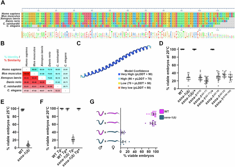
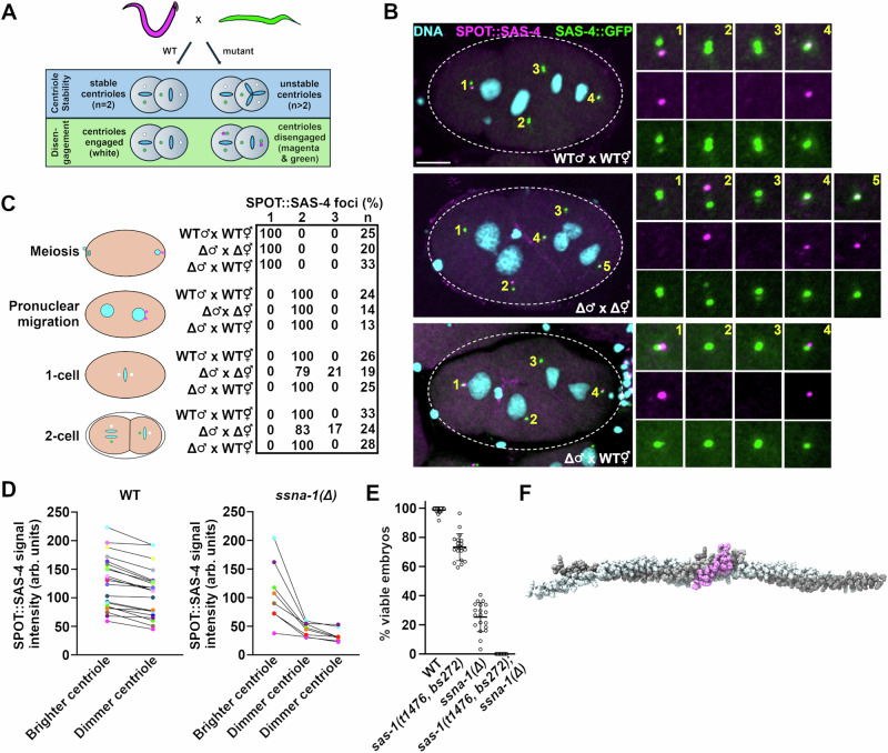
Centrioles play key roles in mitotic spindle assembly. Once assembled, centrioles exhibit long-term stability, but how stability is achieved and how it is regulated are not completely understood. In this study we show that SSNA-1, the Caenorhabditis elegans ortholog of Sjogren's Syndrome Nuclear Antigen 1, is a constituent of centrioles and centriole satellite-like structures. A deletion of ssna-1 results in the formation of extra centrioles. We show that SSNA-1 genetically interacts with the centriole stability factor SAS-1 and is required post assembly for centriole structural integrity. In SSNA-1's absence, centrioles assemble but fracture leading to extra spindle poles. However, if the efficiency of cartwheel assembly is reduced, the absence of SSNA-1 results in daughter centriole loss and monopolar spindles, indicating that the cartwheel and SSNA-1 cooperate to stabilize centrioles during assembly. Our work thus shows that SSNA-1 contributes to centriole stability during and after assembly, thereby ensuring proper centriole number.


Conflict of interest statement

Competing interests: The authors declare no competing interests.

Figures

Fig. 1
Fig. 1. C. elegans possesses an SSNA-1 ortholog that plays a critical role in embryogenesis.
A Sequence alignment of various SSNA-1 orthologs. The amino acids are colored as follows: blue and red for negatively and positively charged residues, respectively, green for uncharged residues, and gold for hydrophobic residues. B Table showing percent identity (blue) and percent similarity (red) of SSNA-1 orthologs. C AlphaFold structural prediction of T07A9.13. D Embryonic viability of wild-type and ssna-1 mutant strains at 20 °C. Each datapoint represents progeny of a single hermaphrodite. Mean and SD are shown. n = 21 (WT), 12 (ssna-1(Δ)), 14 (ssna-1(Δ)/+), 14 (ssna-1G7X), 21 (ssna-1Δ2-17), 12 (ssna-1Δ2-17/+), 21 (ssna-1Δ2-18), 13 (ssna-1Δ2-22), 14 (ssna-1Δ100-105). E Embryonic viability of wild-type and ssna-1(Δ) strains at 25 °C. Each datapoint represents progeny of a single hermaphrodite. Mean and SD are shown. n = 14(WT), 14(ssna-1(Δ)). F Embryonic viability of wild-type and ssna-1(Δ) strains with or without the ssna-1 transgene (Tg). Note that when expressed in the wild type, the transgene does not affect viability, but when expressed in the ssna-1(Δ) mutant it fully rescues the embryonic lethal phenotype. Each datapoint represents progeny of a single hermaphrodite. Mean and SD are shown. n = 13 (WT Tg−), 13 (WT Tg+), 12 (ssna-1(Δ) Tg−), 13 (ssna-1(Δ) Tg+). G SSNA-1 is maternally required. Embryonic viability among the progeny of wild-type (magenta) or ssna-1(Δ) (blue) males and wild-type or ssna-1(Δ), fem-1(hc17) females. Note that embryonic lethality correlates with the genotype of the mother and not the father. No significant differences were observed in viability among the progeny sired by wild-type or mutant males. Each datapoint represents progeny of a single female. Mean and SD are shown. n = 18 (WT × WT), 17 (ssna-1(Δ) males × WT), 10 (WT × ssna-1(Δ) females), 9 (ssna-1(Δ) × ssna-1(Δ)). Source data are provided as a Source Data file.
Fig. 2
Fig. 2. SSNA-1 is a component of the centriole and centriole satellite-like structures.
A Immunofluorescence images of SSNA-1::SPOT and SAS-4::GFP during the first embryonic division. SSNA-1 is found only at centrioles from meiosis through anaphase of the first cell cycle. During first telophase, additional weaker SSNA-1::SPOT foci can be seen (arrowheads). Scale bar = 10 μm and applies to (A and B). Enlargements are 2× magnifications. Experiment was repeated three times with identical results. B During the second cell cycle SSNA-1::SPOT remains centriolar but beginning around nuclear envelope breakdown (NEBD), SSNA-1::SPOT assembles into satellite-like structures that surround the centriole. Enlargements are 2× magnifications. Experiment was repeated three times with identical results. C, D Immunofluorescence image showing co-localization of SSNA-1::SPOT relative to the PCM marker GFP::SPD-5. Twenty-six two-cell embryos were examined with identical results. C During anaphase, SSNA-1::SPOT satellite-like structures localize outside of the PCM marked by GFP::SPD-5. Scale bar = 10 μm and applies to (C and D). D During telophase when the PCM breaks up into packets, SSNA-1::SPOT satellite-like structures remain distinct from SPD-5 packets. However, a portion of both SSNA-1::SPOT and GFP::SPD-5 co-localize at the centriole (arrowheads in enlargements). E Frames from a time-lapse recording of an embryo expressing SSNA-1::wrmScarlet, γ-tubulin::GFP, and GFP::Histone H2B. First and last frame are at top. SSNA-1::wrmScarlet reorganizes during division of the ABpl cell (boxed). Time-lapse images reveals that the satellite-like structures disperse over the nucleus during interphase and accumulate at centrosomes as cells enter mitosis. Time is shown as mm:ss. Scale bar = 10 μm. A total of 40 embryos were imaged with similar results.
Fig. 3
Fig. 3. Centriolar SSNA-1 localizes between the microtubule-containing outer wall and the cartwheel.
AC Ultrastructure Expansion Microscopy (U-ExM) of centrioles from gonad spreads. The schematic shown to the right of each set of images depicts the orientation and configuration of centrioles. A Co-localization of SSNA-1, HA::SAS-4, and α/β-tubulin from two distinct centrioles showing a top view (centriole #1) and side view (centriole #2). SSNA-1 is localized inside the microtubule outer wall. Arrowhead indicates emergence of a daughter centriole that stains weakly for HA::SAS-4 but not SSNA-1. Scale bars = 100 nm. B Co-localization of SSNA-1, SAS-6::HA, and α/β-tubulin. SSNA-1 is localized outside of the cartwheel defined by SAS-6::HA. Arrowhead indicates emergence of a daughter centriole that stains positive for SAS-6::HA but not SSNA-1. Scale bar = 100 nm. C Co-localization of SSNA-1 and HA::ZYG-1. Scale bar = 200 nm. D The measured diameters of each protein are plotted with each point representing a measurement from a single centriole. The mean and standard deviation are shown. n = 28 (SAS-6::HA), 32 (SSNA-1), 55 (α/β-Tubulin), 27 (HA::SAS-4), and 13 (HA::ZYG-1). E Diagram indicating the position of each protein within the centriole. SSNA-1 localizes between SAS-6::HA and α/β-tubulin. F Co-localization of SSNA-1 and SAS-1::3 × Flag in centrioles from gonad spreads. Scale bar = 100 nm. To the right is a signal intensity profile plot of SSNA-1 and SAS-1::3 × Flag. G Co-localization of SSNA-1 and SAS-1::3 × Flag in embryonic centrioles. Scare bar = 100 nm. Source data are provided as a Source Data file.
Fig. 4
Fig. 4. Deletion of SSNA-1 results in excess centrioles.
A Time-lapse images of the first four rounds of division of either wild-type or ssna-1(Δ) embryos expressing GFP::histone, mCherry::β-tubulin, and GFP::SPD-2. While wild-type embryos form only bipolar spindles, ssna-1(Δ) embryos form multipolar spindles beginning around the four-cell stage. During the third round of division in the ssna-1(Δ) embryo (t = 27:00), multiple centrosomes highlighted by cyan, yellow, and white arrowheads appear. These form a multipolar spindle in the EMS cell (t = 33:00). Each centrosome is capable of duplication as shown in the subsequent daughter cells E (white arrowheads) and MS (cyan and yellow arrowheads). Scale bar = 10. B Percent of embryos displaying a multipolar spindle or detached centrosome defect. (n = number of embryos scored). C Percent of embryos at each cell stage displaying at least one multipolar spindle or a detached centrosome defect. The percent of embryos displaying no defect (bipolar spindles only) are represented by black bars. (n = number of embryos scored). D Percentage of tripolar and tetrapolar spindles among all multipolar spindles (n = spindles scored). E Percentage of cells with a bi-, tri- or tetrapolar spindle at each embryonic cell stage (n = number of cells scored). F An immunofluorescent image of a four-cell stage ssna-1(Δ) embryo with a multipolar spindle in both the ABp (1–3) and EMS (4–6) cells. Note that all spindle poles are positive for both ZYG-1::SPOT and SAS-4 indicating the extra spindle poles contain centrioles. Scale bar = 10 μm. Nineteen embryos with multipolar spindles were examined with all poles of all multipolar spindles staining positive for each marker. Source data are provided as a Source Data file.
Fig. 5
Fig. 5. SSNA-1 functions during centriole assembly.
A Embryonic viability of progeny from wild type, zyg-1(it25), ssna-1(Δ), and zyg-1(i25); ssna-1(Δ) strains at 22.5 °C. Each data point represents the progeny of a single hermaphrodite. Mean and SD are shown. n = 10 (WT), 10 (zyg-1(it25)), 10 (ssna-1(Δ)), 10 (zyg-1(it25); ssna-1(Δ)). ***p = 0.0003, ****p < 0.0001 as determined with a one-way ANOVA with Tukey’s multiple comparisons test. B Single frames taken from time-lapse recordings of the indicated strains expressing GFP::histone and SPD-2::mCherry. Each image shows a two-cell stage embryo. A few zyg-1(it25) embryos exhibit monopolar spindles (magenta arrowhead), while ssna-1(Δ) embryos only possess bipolar spindles (white arrowheads). The zyg-1(it25); ssna-1(Δ) double mutant embryos exhibit a mixed phenotype, with monopolar spindles (magenta arrowheads) or tripolar spindles (cyan arrowheads). C Quantification of spindle defects observed through the first two cell divisions at 22.5 °C. The zyg-1(it25); ssna-1(Δ) double mutant embryos display twice as many monopolar spindles as zyg-1(it25) embryos. The double mutant also assembles multipolar spindles which are not observed in either single mutant. D Co-sedimentation assay showing SSNA-1 and ZYG-1 interact. Proteins were incubated alone (left) or in various combinations (right), separated into soluble (S) and pellet (P) fractions by centrifugation, and then detected by immunoblotting. Note that ZYG-1 is found in the soluble fraction when incubated alone or with microtubules but shifts to the pellet fraction upon incubation with SSNA-1. E Quantitation from three-independent co-sedimentation experiments. Mean and SD are shown. Source data are provided as a Source Data file.
Fig. 6
Fig. 6. Extra centrioles arise early during the cell cycle in ssna-1(Δ) embryos.
Time lapse images from wild-type or ssna-1(Δ) embryos expressing mCherry::histone and GFP::SAS-7. The first and last frames are shown at the top while enlargements show intervening time points with only one spindle pole shown beginning in anaphase (time point 3:00). In wild-type embryos two GFP::SAS-7 foci (corresponding to a disengaged centriole pair) first become visible at each spindle pole during telophase (t = 9:00, yellow and magenta arrowheads). During the ensuing cell cycle, this cell assembles a bipolar spindle (t = 18:00). In ssna-1(Δ) embryos, two GFP::SAS-7 foci first become apparent during prometaphase (t = 1.0, yellow and magenta arrowheads). During telophase when centriole separation normally occurs, a third GFP::SAS-7 spot appears (t = 8.0, cyan arrowhead). During the ensuring cell cycle this cell assembles a tripolar spindle (t = 17:00). Bar = 10 μm.
Fig. 7
Fig. 7. SSNA-1 is required for the structural integrity of centrioles.
A Assay to detect centriole fragmentation and premature disengagement. Males whose centrioles are marked with SPOT::SAS-4 (magenta) are mated to hermaphrodites expressing SAS-4::GFP (green). In wild-type embryos the two paternal centrioles will indelibly be marked magenta while new centrioles will be marked green. Magenta mother/green daughter centriole pairs will remain engaged (and appear white) until telophase. Centriole instability will be detected as >2 magenta centrioles per embryo while premature disengagement will present as loss of green-red coincidence prior to telophase. B WT × WT (top), ssna-1(Δ) × ssna-1(Δ) (middle), and ssna-1(Δ) × WT two-cell stage embryos stained for SPOT::SAS-4 (magenta), SAS-4::GFP (green), and DNA (cyan). The WT × WT and ssna-1(Δ) × WT embryos possess two magenta centrioles indicating centriole stability while the ssna-1(Δ) embryo possesses three magenta centrioles revealing a centriole fragmentation phenotype. Note that in the wild-type embryo, one of the blastomeres in telophase is undergoing centriole duplication where one of the sperm centrioles (1) is producing a new green daughter. Bar = 10 μm. C Quantitation of centriole fragmentation in wild-type and ssna-1(Δ) mutant embryos. Note that in early prophase ssna-1(Δ) mutant embryos have two sperm derived centrioles indicating ssna-1(Δ) sperm contain the normal number of centrioles. However, older ssna-1(Δ) embryos frequently contain more than two magenta foci indicating that the sperm centrioles have fragmented. D Quantitation of SPOT::SAS-4 intensities. Lines connect centrioles from the same embryo. In ssna-1(Δ) embryos with three sperm derived centrioles, one is always significantly brighter than the other two, while in wild-type embryos, the sperm centrioles are much more similar in intensity. E ssna-1 and sas-1 genetically interact. The percentage of viable embryos produced by wild-type n = 23, ssna-1(Δ) n = 19, sas-1(t1476, bs272) n = 22, and sas-1(t1476, bs272); ssna-1(Δ) n = 21 hermaphrodites. Each datapoint represents the progeny of a single hermaphrodite. Mean and SD are shown. F AlphaFold prediction of SSNA-1 tetramer (grey) and SAS-1 (a.a. 539–570) (pink) showing an interaction with a pTM score of 0.65. The SSNA-1 tetramer structure is from Agostini et al. Source data are provided as a Source Data file.
Fig. 8
Fig. 8. SSNA-1 functions to stabilize centrioles during and after assembly.
A model depicting the fate of centrioles possessing or lacking stabilizing elements. Top. Wild-type centrioles, which possess both SSNA-1 (yellow ring) and a fully formed cartwheel, exhibit long-term stability resulting in faithful bipolar spindle assembly. Middle. Centrioles lacking SSNA-1 can be assembled but lack long-term stability leading to centriole fragmentation and multipolar spindle formation. Bottom. Centrioles lacking SSNA-1 and a fully formed cartwheel (due to partial inhibition of zyg-1) experience structural failure during assembly leading to loss of the daughter centriole and monopolar spindle assembly. Microtubules are in purple, SSNA-1 is in yellow, and the cartwheel is in black. The green cylinder highlights the lumen of the centriole.

Update of

Similar articles

Cited by

References

    1. Breslow, D. K. & Holland, A. J. Mechanism and regulation of centriole and cilium biogenesis. Annu. Rev. Biochem.88, 691–724 (2019). - PMC - PubMed
    1. Izquierdo, D., Wang, W. J., Uryu, K. & Tsou, M. F. Stabilization of cartwheel-less centrioles for duplication requires CEP295-mediated centriole-to-centrosome conversion. Cell Rep.8, 957–965 (2014). - PMC - PubMed
    1. Le Guennec, M. et al. A helical inner scaffold provides a structural basis for centriole cohesion. Sci. Adv.6, eaaz4137 (2020). - PMC - PubMed
    1. Hilbert, M. et al. SAS-6 engineering reveals interdependence between cartwheel and microtubules in determining centriole architecture. Nat. Cell Biol.18, 393–403 (2016). - PubMed
    1. Kitagawa, D. et al. Structural basis of the 9-fold symmetry of centrioles. Cell144, 364–375 (2011). - PMC - PubMed

MeSH terms

Substances

LinkOut - more resources