. 2025 Jun 5;16(1):5225.
doi: 10.1038/s41467-025-60426-9.

Ca2+-dependent cytoplasmic and nuclear phosphorylation of STOP1 by CPK21 and CPK23 confers ALMT1-dependent aluminum resistance

Affiliations

Ca2+-dependent cytoplasmic and nuclear phosphorylation of STOP1 by CPK21 and CPK23 confers ALMT1-dependent aluminum resistance

Hongrui Cao et al. Nat Commun. .

Abstract

Calcium (Ca) signalling is critical for plant responses to aluminum (Al) stress, with STOP1-mediated ALMT1 expression playing a crucial role in Arabidopsis Al resistance. However, the specific intracellular Ca2+ sensors responsible for transducing Al signals in this process remain unclear. In this study, we identified CPK21 and CPK23, members of the CPK family, as key regulators promoting STOP1-mediated ALMT1 expression under Al stress, significantly influencing malate exudation from roots to limit Al accumulation in root tips. Al stress triggers rapid Ca2+-dependent accumulation of CPK21 and CPK23 in the plasma membrane, cytoplasm and nucleus of root apical cells. The Al-activated CPK21 and CPK23 subsequently phosphorylate STOP1 in both the cytoplasm and nucleus of root apical cells, stabilizing STOP1 by preventing its interaction with RAE1, ultimately enhancing Al resistance. This entire process is Ca2+-dependent. The study unveils a previously undisclosed regulatory network in which CPKs integrate Al-evoked Ca2+ signals and transcriptional reprogramming through the Ca2+-CPK21/23-STOP1 cascade to effectively respond to and adapt to Al stress in plants.


Conflict of interest statement

Competing interests: The authors declare no competing interests.

Figures
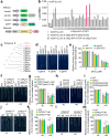
Fig. 1
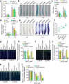
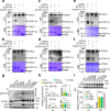
Fig. 1. CPK21/23 promote STOP1-mediated ALMT1 expression and confer plant resistance to Al toxicity.
a Schematics show the construct of STOP1 and CPKac effector and ALMT1p:LUC reporter. b CPKac activates the STOP1-promoted ALMT1p:LUC in Arabidopsis mesophyll protoplasts transient assays. Data represent means ± SD (n = 4 biologically independent samples). The dashed line indicates a two-fold increase in STOP1-promoted ALMT1 promoter activity. The Arabic numerals 1-34 represent CPK1ac-CPK34ac. c Phylogenetic tree of subgroup II CPKs. dm Primary root growth in Al-exposed wild type (WT) plants, mutants (cpk21-1, cpk21-2, cpk23, cpk21/23), complemented lines (CPK21/cpk21-2, CPK23/cpk23), and overexpression lines (CPK21OE and CPK23OE). Seedlings were grown in 2% MGRL solution containing 0, 4 and 6 μM Al (d, e), and 0 or 6 μM Al (fm) at pH 5.0 for 5 days. Data in (e, g, i, k, m) represent means ± SD (e: n = 20 biologically independent plants; g, i, k, m: n = 30 biologically independent plants). Lower-case letters in (e, g, i) indicate significant differences at P < 0.05 in the presence of Al (One-way ANOVA, Tukey-test). Comparisons in (k, m) were performed using unpaired two-tailed t tests (****P < 0.0001).
Fig. 2
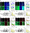
Fig. 2. CPK21/23 promote Al-induced malate exudation, restricting Al accumulation in root tips.
a RT-qPCR analysis of ALMT1 expression in Al-exposed roots of WT and mutants (cpk21-1, cpk23, cpk21/23). UBQ1 was used as a reference gene, and untreated WT plants served as the sample control. Data represent means ± SD (n = 3 biologically independent samples). b, cALMT1p:GUS expression in the root tips of WT and mutants (cpk21-1, cpk23, cpk21/23, stop1) exposed to Al. Bar = 100 μm. Data in (c) represent means ± SD (n = 30 biologically independent plants). d Malate exudation from the roots of WT and mutants (cpk21-1, cpk23, cpk21/23, stop1) in response to Al stress. Data represent means ± SD (n = 4 biologically independent samples). e Hematoxylin staining showing Al accumulation in the roots of WT and mutants (cpk21-1, cpk23, cpk21/23, stop1). Bar = 100 μm. (f) Al content in root tips of WT and mutants (cpk21-1, cpk23, cpk21/23) after exposure to Al. Data represent means ± SD (n = 4 biologically independent samples). Center lines and box edges are medians and the lower and upper quartiles, respectively. Whiskers extend to the lowest and highest data points. For (af), Six-day (ac, e, f) or five-day (d) old seedlings were exposed to 0 and 20 μM Al for 6 h (ac, e, f) or 24 h (d). gl Primary root growth in Al-exposed WT, mutants (cpk21/23 and almt1), overexpression lines (CPK21OE and CPK23OE) and genetically crossed lines (CPK21OE/almt1, CPK23OE/almt1, cpk21/23almt1). Seedlings were grown in 2% MGRL solution containing 0, 2 and 3 μM Al at pH 5.0 for 5 days. Data in (h, j, l) represent means ± SD (n = 30 biologically independent plants). Lower-case letters in (a, c, d, f, h, j, l) indicate significant differences at P < 0.05 in the presence of Al (One-way ANOVA, Tukey-test).
Fig. 3
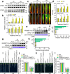
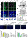
Fig. 3. Al-stimulated CPK21/23 activity is required for Al resistance.
a, b Accumulation of CPK21 and CPK23 in Al-exposed roots of CPK21p:CPK21-3HA or CPK23p:CPK23-3HA transgenic lines. Six-day-old seedlings were exposed to 0 and 20 μM Al at pH 5.0 for 0.5 to 6 h. Data in (b) represent means ± SD (n = 3 biologically independent experiments). c, d Confocal imaging of the CPK21p:CPK21-GFP and CPK23p:CPK23-GFP transgenic lines showing GFP signals in the epidermis of root apical cells. Cell boundaries are defined by red (propidium iodide) staining. Bar = 50 μm. Six-day-old seedlings were exposed to 0 and 20 μM Al at pH 5.0 for 1, 3, or 6 h. Quantitative analysis of mean fluorescence intensity of GFP signals in the epidermis layer of root tips is shown in (d). Data represent means ± SD (n = 10 biologically independent plants). eh In-gel kinase assays showing Al-activated CPK21 and CPK23. Six-day-old seedlings of CPK21p:CPK21-3HA or CPK23p:CPK23-3HA transgenic lines were exposed to either 0 and 20 µM Al at pH 5.0 for 0 to 6 h, and the immunoprecipitated CPK21-HA or CPK23-HA with anti-HA magnetic beads were subjected to in-gel kinase assays. Kinase activity was determined using the p-Thr antibody, with GST-NRAMP1-N as the substrate. Relative kinase activity is shown in (f) and (h), with the intensity at 0 h set to 1.0 (refer to the Methods section for detailed information). Data represent means ± SD (n = 3 biologically independent experiments). (i-l) Primary root growth in Al-exposed WT, mutants (cpk21 and cpk23) and complementation lines (CPK21WT/cpk21-2, CPK21D204A/cpk21-2, CPK23WT/cpk23, CPK23D193A/cpk23). Seedlings were grown in 2% MGRL solution containing 0, 4 and 6 μM Al at pH 5.0 for 5 days. Data in (j, l) represent means ± SD (n = 30 biologically independent plants). Comparisons in (b, d) were performed using unpaired two-tailed t tests (*P < 0.05; **P < 0.01; ***P < 0.001, ****P < 0.0001). Lower-case letters in (f, h, j, l) indicate significant differences at P < 0.05 in the presence of Al (One-way ANOVA, Tukey-test).
Fig. 4
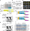
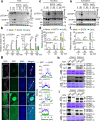
Fig. 4. CPK21/23 interact with STOP1.
a Yeast two-hybrid assay. Yeast cells were grown on SD/-Leu/-Trp (SD-LW) medium or SD/-Leu/-Trp/-His/-Ade (SD-LWHA) medium. X-α-Gal was directly spotted onto the yeast colonies. b, c Co-IP assay. Constructs harboring GFP-CPK21/STOP1-MYC or GFP-CPK23/STOP1-MYC were transformed into N. benthamiana leaves and expressed for 48 h at 22 oC. Total proteins were extracted and incubated with an anti-GFP antibody. The immunoprecipitated proteins were detected with an anti-MYC and anti-GFP antibody. d LCA assay. Constructs harboring cLUC-STOP1/CPK21-nLUC or cLUC-STOP1/CPK23-nLUC were transformed into N. benthamiana leaves and expressed for 48 h at 22 oC. nL: nLUC; cL: cLUC. e Schematic diagram of STOP1 used for the GST pull-down assay in (f). f, g Pull-down assay showing the interaction of CPK21 and CPK23 with STOP1-N and STOP1-C. Input and output were analyzed via Western blot using anti-MBP and anti-GST antibodies. Arrows indicate the desired protein. h Schematic diagram of CPK21 and CPK23 used for yeast two-hybrid assay in (i). i Yeast two-hybrid assays showing the interaction of CPK21 and CPK23 protein fragments with STOP1. Yeast cells were grown on SD/-Leu/-Trp (SD-LW) medium or SD/-Leu/-Trp/-His/-Ade (SD-LWHA) medium. X-α-Gal was directly spotted onto the yeast colonies. The experiment in (b, c, f, g) was repeated 3 times with similar results.
Fig. 5
Fig. 5. CPK21/23 phosphorylate STOP1 and affect its stability.
a λPPase treatment abolishes Al-induced Phos-tag gel shift of STOP1. Proteins were extracted from STOP1p:STOP1-3HA transgenic roots treated with or without 20 μM Al for 1 h, followed by λPPase treatment for 30 min before Phos-tag analysis. Upper bands in the Phos-tag gel represent phosphorylated STOP1 (pSTOP1). b, c STOP1 phosphorylation and accumulation in Al-exposed Wild type (STOP1p:STOP1-3HA), mutants (cpk21/STOP1p:STOP1-3HA, cpk23/STOP1p:STOP1-3HA) and overexpression lines (CPK21/STOP1p:STOP1-3HA, CPK23/STOP1p:STOP1-3HA) were exposed to 0 and 20 μM Al at pH 5.0 for 3 h. Extracted proteins were analyzed using Phos-tag and standard western blotting. Upper bands in the Phos-tag gel indicate phosphorylated STOP1, the middle panel shows the total STOP1 protein level detected with anti-HA antibody, and the lower panel shows the internal reference Anti-Actin. d CPK21 and CPK23 phosphorylate STOP1 in vitro. Purified recombinant proteins of MBP (empty vector), MBP-CPK21, or MBP-CPK23 were incubated with STOP1-6His. Phosphorylated STOP1 was detected using anti-pThr and anti-pSer antibodies. e, f CPK21 and CPK23 kinase dead mutants failed to phosphorylate STOP1 in vitro. Purified recombinant proteins of MBP, MBP-CPK21, MBP-CPK21D204A, MBP-CPK23, or MBP-CPK23D193A were incubated with STOP1-6His. Phosphorylated STOP1 was detected using anti-pThr and anti-pSer antibodies. g, h STOP1p:STOP1-GUS activity analysis in Al-exposed root tips of WT and mutants (cpk21, cpk23, cpk21/23). Bar = 100 μm. i, j Confocal imaging shows reduced nuclear accumulation of STOP1 in Al-exposed root tips of WT and mutants (cpk21, cpk23, cpk21/23). Cell boundaries are defined by red (propidium iodide) staining. Bar = 50 μm. Data in (h, j) represent means ± SD (n = 40 biologically independent plants). Five-day-old STOP1p:STOP1-GUS (g, h) or STOP1p:STOP1-GFP (i, j) transgenic seedlings in WT and mutants were exposed to 0 and 20 μM Al at pH 5.0 for 6 h. Lower-case letters in (h, j) indicate significant differences at P < 0.05 in the presence of Al (One-way ANOVA, Tukey-test). In (j): The thick dashed lines of violin plots represent the median, while thin dashed lines represent the first or third quartile. The experiment in (af) was repeated 3 times with similar results.
Fig. 6
Fig. 6. The residues Thr31, Ser163, and Thr316 are key sites for CPK21- and CPK23-mediated phosphorylation and the stability of STOP1.
af Phospho-null mutations of Thr31, Ser163 and Thr316 strongly suppressed CPK21/23-mediated phosphorylation of STOP1 in vitro. Purified recombinant proteins of MBP-CPK21 or MBP-CPK23 were incubated with GST-STOP1-N, GST-STOP1-NT31A, GST-STOP1-NS163A, GST-STOP1-C, or GST-STOP1-CT316A. Phosphorylated STOP1 were detected using anti-pThr or anti-pSer antibodies. g, h Phospho-null mutations of Thr31, Ser163 and Thr316 significantly affect the phosphorylation and stability of STOP1 in vivo. i, j Effect of phosphor-mimetic mutations of Thr31, Ser163 and Thr316 on STOP1 stability in vivo. Six-day-old seedlings of WT (STOP1p:STOP1WT-GFP/stop1), cpk21/23 mutant (cpk21/23/STOP1p:STOP1-GFP), phospho-null mutants (STOP1p:STOP1T31A-GFP/stop1 STOP1p:STOP1S163A-GFP/stop1, STOP1p:STOP1T316A-GFP/stop1) (g, h), and phosphor-mimetic mutants (STOP1p:STOP1T31D-GFP/stop1, STOP1p:STOP1S163D-GFP/stop1, STOP1p:STOP1T316D-GFP/stop1) (i, j) were exposed to 0 and 20 μM Al at pH 5.0 for 3 h. Extracted proteins were analyzed using Phos-tag and standard western blotting. In (g, h): Upper bands in the Phos-tag gel indicate phosphorylated STOP1, the middle panel shows the total STOP1 protein level detected with anti-GFP antibody, and the lower panel shows the internal reference, Anti-Actin. Relative phosphorylated and total STOP1 levels in (g) are shown in (h), and relative total STOP1 levels in (i) are shown in (j). Data in (h, j) represent means ± SD (n = 3 biologically independent experiments). Comparisons in (h, j) were performed using unpaired two-tailed t tests (**P < 0.01; ***P < 0.001, ****P < 0.0001). The experiment in (af) was repeated 3 times with similar results.
Fig. 7
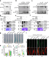
Fig. 7. Al enhances the accumulation of CPK21 and CPK23 in the plasma membrane, cytoplasm and nucleus.
a, b, j, h Confocal imaging analysis showing the cellular localization of CPK21-GFP and CPK23-GFP in root apical cells under Al stress. Six-day-old seedlings of transgenic lines expressing CPK21p:CPK21-GFP and CPK23p:CPK23-GFP were exposed to 0 and 20 μM Al at pH 5.0 for 6 h. DAPI and FM4-64 signals indicate the nucleus and plasma membrane localization, respectively. Scale bars: 50 μm (long solid line) and 5 μm (short solid line). cf, il Immunoblot analysis of cellular CPK21 and CPK23 localization in Al-exposed roots. CPK21-GFP and CPK23-GFP were detected using anti-GFP antibodies (c, e), while CPK21-HA and CPK23-HA were detected with anti-HA antibodies (i, k). UDP-glucose pyrophosphorylase (UGPase) (c, e, i, k), histone H3 (H3) (c, e) and H+-ATPase (i, k) were used as loading controls for cytosolic, nuclear and plasma membrane fractions, respectively. In (d, f), the relative protein levels of CPK21 (c) and CPK23 (e) in total, cytoplasmic, and nuclear fractions were normalized to their respective markers, yielding RT, RC, and RN values. The ratio RT(-Al)/RT(-Al) was set to as Rref (1.0), and the relative protein levels were calculated as RT/RT(-Al), RC/RT(-Al), and RN/RT(-Al). Similarly, in (j, l), the levels of CPK21 (i) and CPK23 (k) in total, plasma membrane, and cytoplasmic fractions were normalized to their relevant markers. RT(-Al)/RT(-Al) was also set as Rref (1.0), with relative protein levels calculated accordingly. T: Total; C: Cytoplasm; N: Nucleus; PM: Plasma Membrane. Data in (d, f, j, l) represent means ± SD (n = 3 biologically independent experiments). Comparisons in (d, f, j, l) were performed using unpaired two-tailed t tests (**P < 0.01; ***P < 0.001, ****P < 0.0001). The experiment in (a, b, g, h) was repeated 3 times with similar results.
Fig. 8
Fig. 8. Residues Thr31, Ser163 and Thr316 are crucial for the cytoplastic and nuclear accumulation and phosphorylation of STOP1, influencing Al resistance.
ad Phospho-null mutations at Thr31, Ser163 and Thr316 significantly reduce STOP1 accumulation and phosphorylation in both the cytoplasm and nucleus of Al-exposed root apical cells. a shows representative images of STOP1-GFP and quantitative analysis in WT plants, cpk21/23 mutants and phosphor-null mutants. DAPI staining marks the nucleus. Scale bars: 50 μm (long solid line) and 5 μm (short solid line). bd display Phos-tag analysis of STOP1 phosphorylation in nuclear (b) and cytoplasmic (c) fractions. In (b, c): Upper bands in the Phos-tag gel indicate phosphorylated STOP1, the middle panel shows the total STOP1 protein level detected with anti-GFP antibody, and the lower panel shows the internal reference Anti-H3 and Anti-UGPase, respectively. Relative phosphorylated STOP1 levels in (b, c) are shown in (d), and relative total STOP1 levels in (b, c) are shown in Supplementary Fig. 23. For quantitative analysis of phosphorylated STOP1 levels in nucleus (b) and cytoplasm (c), the amount of phosphorylated STOP1 in nuclear and cytoplasmic fractions was normalized to relevant markers (RC and RN, respectively), with RN or C(STOP1WT/-Al)/RN or C(STOP1WT/-Al) set as Rref (1.0). Relative protein levels were calculated as RN/RN(STOP1WT/-Al) and RC/RC(STOP1WT/-Al). Data in (d) represent means ± SD (n = 3 biologically independent experiments); comparisons were performed using unpaired two-tailed t tests (***P < 0.001, ****P < 0.0001). eh Primary root growth in Al-exposed WT, stop1 mutant, and complementary lines expressing STOP1, phospho-null STOP1 mutations (STOP1T31A/stop1, STOP1S163A/stop1 and STOP1T316A/stop1), or phosphor-mimetic mutations (STOP1T31D/stop1, STOP1S163D/stop1 and STOP1T316D/stop1). Seedlings were grown in 2% MGRL solution containing 0, 2 and 3 μM (e, f) or 0 and 6 μM Al (g, h) (pH 5.0) for 5 days. Data in (f, h) represent means ± SD (n = 30 biologically independent plants); significant differences (P < 0.05) in the presence of Al were indicated by different letters (One-way ANOVA, Tukey-test).
Fig. 9
Fig. 9. CPK21/23-mediated phosphorylation and stability of STOP1 under Al stress is Ca2+-dependent.
af Immunoblots and Phos-tag analysis of STOP1 abundance and phosphorylation in Al-exposed roots (a, b) or in nuclear (c, d) and cytoplasmic (e, f) fractions of Al-exposed root cells with or without EGTA and LaCl3 treatment. Six-day-old STOP1p:STOP1-3HA transgenic seedlings were pretreated with or without 20 mM EGTA for 1 h or 2 mM LaCl3 for 20 min, followed by treatment with 2% MGRL solution containing 0 and 20 μM Al (pH 5.0) for 60 min. Proteins were separated on 6% phos-tag-PAGE and 12% SDS-PAGE and detected using anti-HA antibody. Histone H3 (H3) (c) and UDP-glucose pyrophosphorylase (UGPase) (e) were used as loading controls for nuclear and cytoplasmic fractions, respectively. Data in (b, d, f) represent means ± SD (n = 3 biologically independent experiments); Comparisons were performed using unpaired two-tailed t tests (***P < 0.001, ****P < 0.0001, ns: not significant). g Confocal imaging analysis showing representative images of STOP1-GFP in Al-exposed root tips, with or without EGTA treatment. Six-day-old STOP1p:STOP1-GFP transgenic seedlings were pretreated with or without 20 mM EGTA for 1 h, followed by exposure to 2% MGRL solution containing 0 and 20 Al μM Al (pH 5.0) for 3 h. Quantified GFP fluorescence intensity is shown on the right. DAPI staining indicates nuclear localization. C: cytoplasm; N: nucleus. Scale bars: 50 μm (long solid line) and 5 μm (short solid line). (h, i) Phosphorylation of STOP1 by CPK21 and CPK23 is Ca2+-dependent. Phosphorylated STOP1 was visualized by western blotting using anti-pThr antibody (Upper panel). Recombinant proteins used in the reaction are shown by Coomassie brilliant blue (CBB) staining (lower panel). STOP1-N: N-terminal of STOP1; STOP1-C: C-terminal of STOP1. The experiment in (gi) was repeated 3 times with similar results.
Fig. 10
Fig. 10. Schematic representation of the Al-Ca2+-CPK21/23-STOP1 signaling cascade involved in ALMT1-dependent Al resistance.
Al induces a rapid accumulation of CPK21 and CPK23 in the plasma membrane, cytosol and nucleus of root apical cells, where they perceive the Al-evoked cytosolic Ca2+ signal. Activated CPK21 and CPK23 subsequently phosphorylate STOP1 in both the cytoplasm and nucleus, stabilizing STOP1 by inhibiting its interaction with RAE1, ultimately enhancing Al resistance through mediating the expression of ALMT1 in the nucleus.

Similar articles

References

    1. von Uexkull, H. R. & Mutert, E. Global extent, development and economic-impact of acid soils. Plant Soil171, 1–15 (1995).
    1. Horst, W. J., Asher, C. J., Cakmak, J., Szulkiewicz, P. & Wissemeier, A. H. Short-term responses of soybean roots to aluminium. J. Plant Physiol.140, 174–178 (1992).
    1. Delhaize, E. & Ryan, P. R. Aluminum toxicity and tolerance in plants. Plant Physiol.107, 315–321 (1995). - PMC - PubMed
    1. Kochian, L. V., Pineros, M. A., Liu, J. & Magalhaes, J. V. Plant adaptation to acid soils: the molecular basis for crop aluminum resistance. Annu. Rev. Plant Biol.66, 571–598 (2015). - PubMed
    1. Ma, J. F., Ryan, P. R. & Delhaize, E. Aluminium tolerance in plants and the complexing role of organic acids. Trends Plant Sci.6, 273–278 (2001). - PubMed

MeSH terms

LinkOut - more resources