. 2025 Jun 5;16(1):5242.
doi: 10.1038/s41467-025-60516-8.

Mechanism of human α3β GlyR regulation by intracellular M3/M4 loop phosphorylation and 2,6-di-tert-butylphenol interaction

Affiliations

Mechanism of human α3β GlyR regulation by intracellular M3/M4 loop phosphorylation and 2,6-di-tert-butylphenol interaction

Xiaofen Liu et al. Nat Commun. .

Abstract

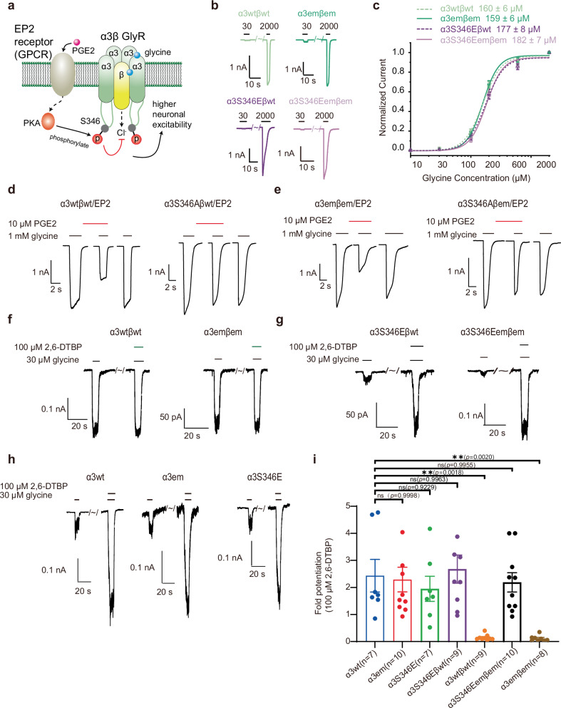
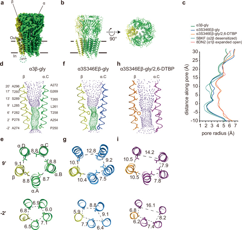
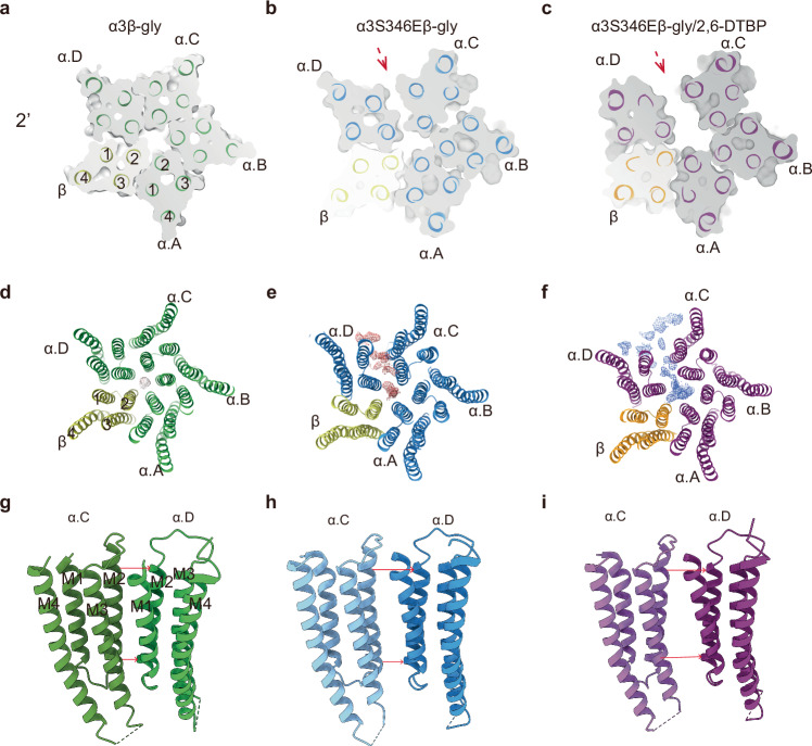
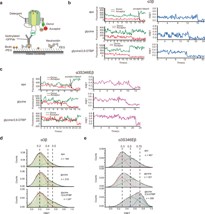
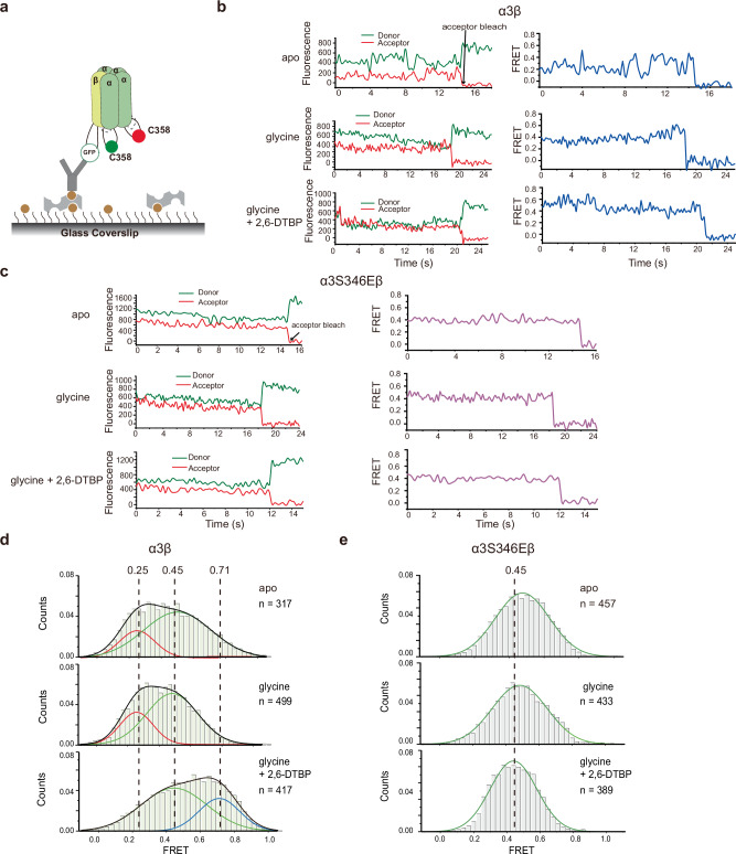
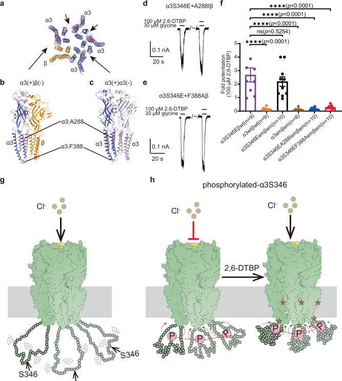
α3β glycine receptor (GlyR) is a subtype of GlyRs that belongs to the Cys-loop receptor superfamily. It is highly expressed in the spinal dorsal horn where sensory information is integrated. Under inflammatory conditions, the large unstructured intracellular M3/M4 loops of the α3 subunit are phosphorylated through the prostaglandin E2 (PGE2) pathway, inhibiting ion conduction, and resulting in elevated pain sensation. A small molecule analgesic analog, 2,6-di-tert-butylphenol (2,6-DTBP) potentiates phosphorylated α3β GlyR through unclear mechanisms and relieves pain. Combining cryo-Electron Microscopy (cryo-EM) structures and single molecule Förster resonance energy transfer (smFRET) experiments, we show compaction of M3/M4 loop towards the ion conduction pore upon phosphorylation and further by 2,6-DTBP binding, which in turn modulates function through changing pore conformations and local electrostatics. We show that simultaneous interactions with the M3/M4 loop and the transmembrane domain (TM) is necessary for the potentiation of heteromeric α3β GlyR by 2,6-DTBP, while TM interaction alone is sufficient to potentiate homomeric α3 GlyR, explaining the mystery of why 2,6-DTBP potentiates only phosphorylated α3β GlyR. These findings show how post-translational modification of the unstructured intracellular M3/M4 loop may regulate Cys-loop receptor function, providing new perspectives in pain control and other pharmaceutical development targeting GlyRs and other Cys-loop receptors.


Conflict of interest statement

Competing interests: The authors declare no competing interests.

Figures

Fig. 1
Fig. 1. Functional characterization of human α3β GlyR.
a The effect of PGE2 on glycinergic signaling. PGE2 activates EP2 receptors, which in turn phosphorylate α3 subunits in a PKA-dependent manner and inhibits Cl- flux through α3β GlyR. b Typical glycine response of α3wtβwt, α3emβem, α3S346Eβwt and α3S346Eemβem at 30 µM and 2 mM glycine concentration. c Dose response with Hill fits (lines). Data are represented as mean ± S.E.M. (α3wtβwt: n = 7 cells, α3emβem: n = 7 cells, α3S346Eβwt: n = 11 cells, α3S346Eemβem: n = 7 cells). d, e Representative recordings showing the effect of 10 μM PGE2 on glycine-evoked current in HEK293T cells co-transfected with the EP2 receptor and d α3wtβwt, and e α3emβem. PKA consensus sequence mutation S346A is present in the right panels. f, g, h Representative whole-cell recordings of glycine-induced currents in the absence and presence of 100 μM 2,6-DTBP in HEK293T cells expressing f α3wtβwt (left) and α3emβem (right) GlyRs, g α3S346Eβwt (left) and α3S346Eemβem (right), and h α3wt (left), α3em (middle), α3S346E (right). i Fold-increase in 30 μM glycine-evoked currents of α3wtβwt, α3emβem, α3S346Eβwt, α3S346Eemβem, α3wt, α3em and α3S346E by 100 μM 2,6-DTBP. Data are represented as mean ± S.E.M. P-values were calculated using one-way ANOVA. (**p  <  0.01; ns, not significant. The exact p-values are shown in the figure).
Fig. 2
Fig. 2. Conformational differences between α3β-gly, α3S346Eβ-gly, and α3S346Eβ-gly/2,6-DTBP GlyRs.
a Side view of the cryo-EM map of α3β GlyR in digitonin in the presence of glycine. b Side (left) and top-down (right) view of the atomic models. α3 subunits and β subunit are respectively colored in bright green and yellow. c Plot of pore radii calculated by the HOLE program for the α3β-gly (green), α3S346Eβ-gly (blue), α3S346Eβ-gly/2,6-DTBP (orange), α2β-gly (desensitized, PDB ID: 5BKF, green dashed line), and α1β-gly (expanded open, PDB ID:8DN2, red dashed line). d, f, h Ion permeation pathways for d α3β-gly, f α3S346Eβ-gly, and h α3S346Eβ-gly/2,6-DTBP GlyRs. M2 helices are shown as cartoons, and the side chains of pore-lining residues as sticks. Purple, green, and red spheres define radii of > 3.3 Å, 1.8-3.3 Å, and <1.8 Å, respectively. e, g, i Cross-sections of M2 helices at residues 9’ (top) and -2’ (bottom) for e α3β-gly, g α3S346Eβ-gly, and i α3S346Eβ-gly/2,6-DTBP GlyRs with distances between neighboring Cα shown in Å.
Fig. 3
Fig. 3. Structural changes in the TMD of α3β and α3S346Eβ GlyRs and induced by 2,6-DTBP binding.
Z-slice in the TMD at 2’ for a α3β-gly, b α3S346Eβ-gly, and c α3S346Eβ-gly/2,6-DTBP. d, e, f Top-down view of d α3β-gly,e α3S346Eβ-gly, and f α3S346Eβ-gly/2,6-DTBP. Maps are contoured at 5 RMSD. Unmodeled densities are shown as mesh and colored in brown for α3β-gly, red for α3S346Eβ-gly, and blue for α3S346Eβ-gly/2,6-DTBP, respectively. g, h, i, Cartoon representation of the TMD interfaces between α.C and α.D subunits for g α3β-gly, h α3S346Eβ-gly, and i α3S346Eβ-gly/2,6-DTBP. The red arrows highlight the distances between adjacent α subunits at -2’ (down) and 19’(top).
Fig. 4
Fig. 4. α3S346E mutation and 2,6-DTBP modulate conformation of the internal M3/M4 loop in α3S346Eβ GlyR.
a Schematic of single-molecule FRET measurements of intra-M3/M4 loop changes. b, c Representative fluorescence (Left) and FRET (Right) traces of b α3β GlyR, and c α3S346Eβ GlyR, in apo (top), + glycine (middle) and + glycine + 2,6-DTBP (bottom) conditions. d, e Histograms of FRET values of d α3β and e α3S346Eβ GlyRs in the apo (top), + glycine (middle) and +glycine + 2,6-DTBP (bottom) conditions, deconvoluted by fitting with two Gaussians (red and green lines represent each Gaussian and black line shows their sum).
Fig. 5
Fig. 5. α3S346E mutation and 2,6-DTBP affect the distances of M3/M4 loops between different α3 subunits.
a Schematic of single-molecule FRET measurements between M3/M4 loops from different α3 subunits. b, c Representative fluorescence (Left) and FRET (Right) traces of b α3β GlyR and c α3S346Eβ GlyR in the apo (top), + glycine (middle), and + glycine + 2,6-DTBP (bottom) conditions. d, e Histograms of FRET values of d α3β and e α3S346Eβ GlyRs in the apo (top), + glycine (middle), and + glycine + 2,6-DTBP (bottom) conditions. FRET value histograms were deconvoluted by fitting with two Gaussians (red and green lines represent each Gaussian and black lines show their sum). e α3S346Eβ GlyRs FRET was fitted with a single Gaussian (green lines).
Fig. 6
Fig. 6. Proposed mechanism for α3 M3/M4 loop phosphorylation and 2,6-DTBP modulation of α3β GlyR activity.
a Top view of TM sectioned near A288, with potential 2,6-DTBP sites indicated by arrows. b, c Sideview of b α3(+)β(-) and c α3(-)β(+) interfaces. A288 and F388 of the α3 subunits are shown as spheres. d, e Representative whole-cell recordings of glycine-induced currents from HEK293T cells expressing d α3S346EA288Iβ and e α3S346EF388Aβ GlyRs with and without 100 μM 2,6-DTBP. f Fold-increases of 30 μM glycine-evoked currents by 100 μM 2,6-DTBP. Data are represented as mean ± S.E.M. P-values were calculated using one way ANOVA. (**p  <  0.01; ns, not significant. the exact p-values are shown in the figure). gh Mechanistic model of M3/M4 loop and 2,6-DTBP regulation. g During the entire functional cycle of non-phosphorylated α3β GlyR (close, open, and desensitized states), the M3/M4 loops of α3 subunits are very flexible and far away from the ion conduction pore in TM. h Left: Upon α3S346 phosphorylation, the M3/M4 loop compacts towards the TM, and distances between adjacent M3/M4 loops becomes more stable. These changes result in pore conformational changes and local electronegativity that decrease Cl- conductance. Right: 2,6-DTBP binds to both TM (stars indicates potential binding sites) and phosphorylated M3/M4 loop, leading to further approximation of loop towards the TM, resulting in pore conformational changes that facilitate Cl- conduction.

Update of

Similar articles

References

    1. Lynch, J. W. Molecular structure and function of the glycine receptor chloride channel. Physiol. Rev.84, 1051–1095 (2004). - PubMed
    1. Lynch, J. W. Native glycine receptor subtypes and their physiological roles. Neuropharmacology56, 303–309 (2009). - PubMed
    1. Yu, H., Bai, X. C. & Wang, W. Characterization of the subunit composition and structure of adult human glycine receptors. Neuron109, 2707–2716.e2706 (2021). - PubMed
    1. Zhu, H. & Gouaux, E. Architecture and assembly mechanism of native glycine receptors. Nature599, 513–517 (2021). - PMC - PubMed
    1. Liu, X. & Wang, W. Asymmetric gating of a human hetero-pentameric glycine receptor. Nat. Commun.14, 6377 (2023). - PMC - PubMed

LinkOut - more resources