. 2025 Jun 5;15(1):19776.
doi: 10.1038/s41598-025-04471-w.

Islet delta-cell architecture is remodelled in the human pancreas during type 1 diabetes

Affiliations

Islet delta-cell architecture is remodelled in the human pancreas during type 1 diabetes

Angie Tegehall et al. Sci Rep. .

Abstract

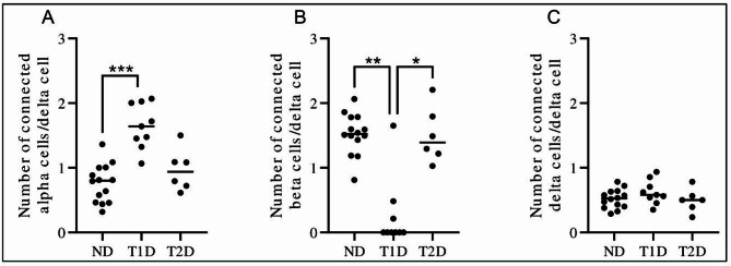
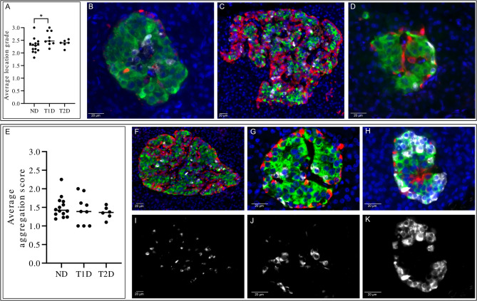
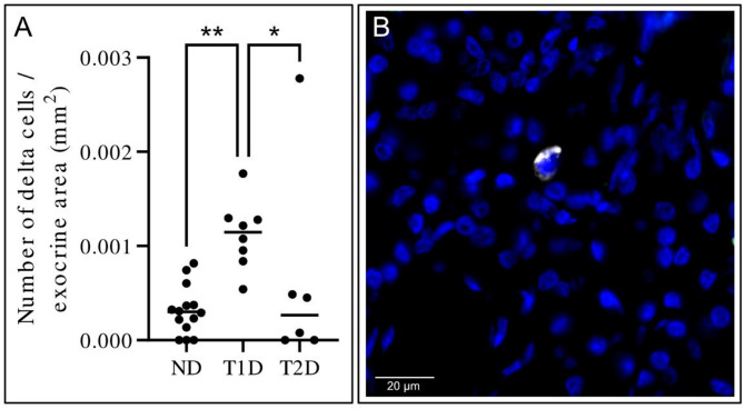
Delta cells participate in regulating hormone secretion in adjacent alpha- and beta cells and a general assumption is that cells with a shorter distance to the secreting cell receive a higher concentration of the secretory compounds. Isolated islets obtained from donors with type 1 diabetes have a reduced glucagon secretion during low glucose levels, but adding a somatostatin receptor inhibitor increases the glucagon secretion. Despite this, information regarding the delta-cell architecture during diabetes is sparse. The aim of the current study was to determine intra-islet and extra-islet delta-cell architecture in the pancreas during long-standing type 1 diabetes or type 2 diabetes. Pancreatic tissue from nine donors with long-standing type 1 diabetes, six donors with type 2 diabetes, and 13 donors without diabetes were obtained. Sections co-stained for somatostatin, glucagon, and insulin were manually examined. There was an approximately two-fold higher number of alpha cells directly adherent to delta cells in subjects with type 1 diabetes compared with non-diabetic subjects. The delta cells were more peripherally located within the islets of donors with type 1 diabetes. The density of extra-islet single delta cells in the acinar region was more than three-fold higher in type 1 diabetes compared with non-diabetic subjects. No differences in delta-cell architecture could be determined in type 2 diabetes compared to non-diabetic subjects. In conclusion, the islet delta-cell architecture in human type 1 diabetes is remodelled. The higher number of alpha cells directly adherent to delta cells in type 1 diabetes likely increases the alpha cells' exposure to somatostatin. This finding may be a link partly explaining the reduced glucagon response to hypoglycemia in type 1 diabetes.

Keywords: Alpha cell; Delta cell; Hypoglycemia; Somatostatin; Type 1 diabetes; Type 2 diabetes.


Conflict of interest statement

Declarations. Competing interests: The authors declare no competing interests.

Figures

Fig. 1
Fig. 1
Average number of alpha, beta or delta cells connected to a delta cell. For each confirmed delta cell, the somatostatin-, glucagon- and insulin-positive cells directly adherent to the somatostatin-positive area were counted. The average number of connected cell types was calculated in each donor for all examined cells in the head, body and tail of the pancreas. (A) Number of connected alpha cells per delta cell, (B) number of connected beta cells per delta cell, and (C) number of connected delta cells per delta cell in non-diabetic (ND), type 1 diabetes (T1D) and type 2 diabetes (T2D) pancreases. Each dot represents an individual donor. The bar represents the median value of the group. *, P < 0.05, **, P < 0.01, ***, P < 0.001.
Fig. 2
Fig. 2
Representative images of islets with immunofluorescent triple-staining of insulin (green), glucagon (red) and somatostatin (white), are shown in a non-diabetic donor (A), a donor with type 1 diabetes (B) and a donor with type 2 diabetes (C). Scale bar: 20 μm.
Fig. 3
Fig. 3
Semi-quantification of delta cell location and aggregation. In each islet it was determined whether delta cells were (1) centrally located, (2) equally distributed throughout the islet or (3) in the peripheral region of the islet. It was also determined whether the islets contained mainly: (1) single delta cells without any connection to other delta cells, (2) pairs of two delta cells, or (3) aggregates of ≥ 3 inter-connected delta cells. (A) The average location grade of all islets in each individual donor was determined in non-diabetic (ND), type 1 diabetes (T1D) and type 2 diabetes (T2D) pancreases. Each dot represents a donor. The bar represents the median value of the group. *, P < 0.05. Representative images of islets, based on immunofluorescent triple-staining of insulin (green), glucagon (red) and somatostatin (white) are shown in B-D. (B) Islet showing mainly centrally located delta cells. (C) Islet with delta cells equally distributed throughout the islet. (D) Islet with delta cells mainly located in the peripheral region of the islet. Scale bar: 20 μm. (E) The average aggregation score of all islets in each individual donor was determined in non-diabetic (ND), type 1 diabetes (T1D) and type 2 diabetes (T2D) pancreases. Each dot represents a donor. The bar represents the median value of the group. There were no statistically significant differences between the groups. Representative images of islets, based on immunofluorescent triple-staining of insulin (green), glucagon (red) and somatostatin (white) are shown in F-K. F, I) Islet with mainly single delta cells without any connection to other delta cells. (F) overlay, I) somatostatin. G, J) Islet with mainly pairs of two delta cells. (G) overlay, J) somatostatin. H, K) Islet with mainly aggregates of ≥ 3 inter-connected delta cells. (H) overlay, K) somatostatin. Scale bar: 20 μm.
Fig. 4
Fig. 4
Number of delta cells per exocrine area (mm2). (A) Single delta cells in the exocrine region of the pancreas were counted in non-diabetic (ND), type 1 diabetes (T1D) and type 2 diabetes (T2D) donor pancreases. The bar represents the median value of the group. Each dot represents a donor. *, P < 0.05, **, P < 0.01. (B) Representative image based on immunofluorescent staining of somatostatin (white) and nuclei (blue) illustrating a single delta cell in the exocrine region of the pancreas. Scale bar: 20 μm.

Similar articles

References

    1. Karimian, N. et al. Somatostatin receptor type 2 antagonism improves glucagon counterregulation in biobreeding diabetic rats. Diabetes62 (8), 2968–2977. 10.2337/db13-0164 (2013). - PMC - PubMed
    1. Kailey, B. et al. SSTR2 is the functionally dominant somatostatin receptor in human pancreatic β- and α-cells. Am. J. Physiol. - Endocrinol. Metab.303 (9), E1107–E1116. 10.1152/ajpendo.00207.2012 (2012). - PMC - PubMed
    1. Gerich, J. E., Langlois, M., Noacco, C., Karam, J. H. & Forsham, P. H. Lack of glucagon response to hypoglycemia in diabetes: evidence for an intrinsic pancreatic alpha cell defect. Science182 (4108), 171–173. 10.1126/science.182.4108.171 (1973). - PubMed
    1. Siafarikas, A. et al. Early loss of the glucagon response to hypoglycemia in adolescents with type 1 diabetes. Diabetes Care. 35 (8), 1757–1762. 10.2337/dc11-2010 (2012). - PMC - PubMed
    1. Singer-Granick, C., Hoffman, R. P., Kerensky, K., Drash, A. L. & Becker, D. J. Glucagon responses to hypoglycemia in children and adolescents with IDDM. Diabetes Care. 11 (8), 643–649. 10.2337/diacare.11.8.643 (1988). - PubMed

MeSH terms

LinkOut - more resources