. 2025 Jun 5;9(1):163.
doi: 10.1038/s41698-025-00964-z.

TRIM65 regulates glucose metabolic reprogramming to promote glioma cell proliferation via ubiquitination and degradation of AMPK

Affiliations

TRIM65 regulates glucose metabolic reprogramming to promote glioma cell proliferation via ubiquitination and degradation of AMPK

Ming-Hui Li et al. NPJ Precis Oncol. .

Abstract

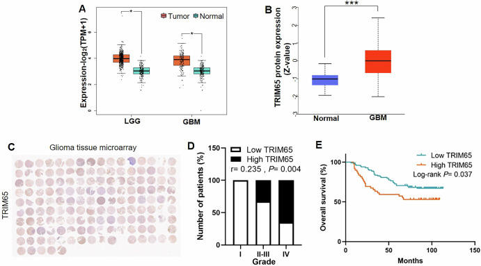
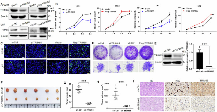
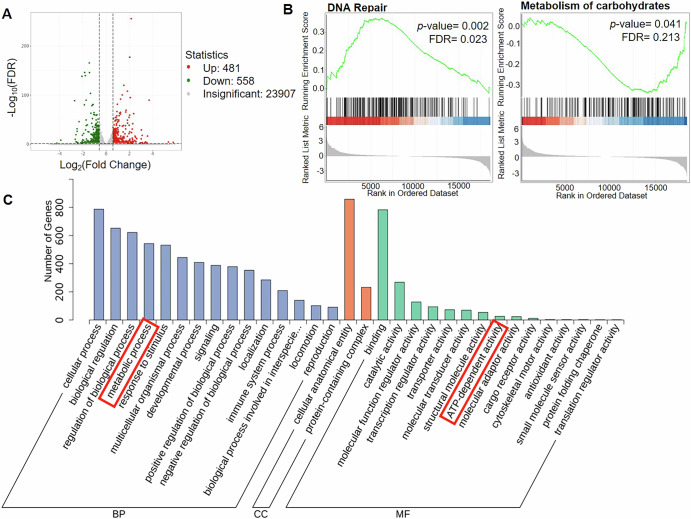
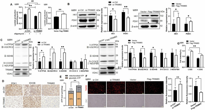
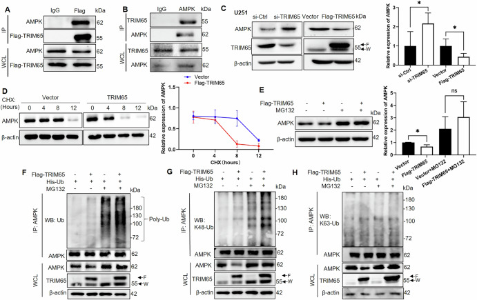
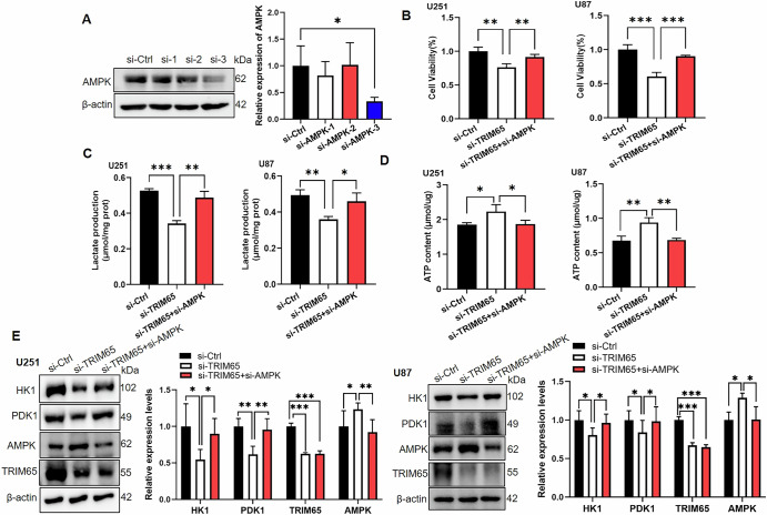
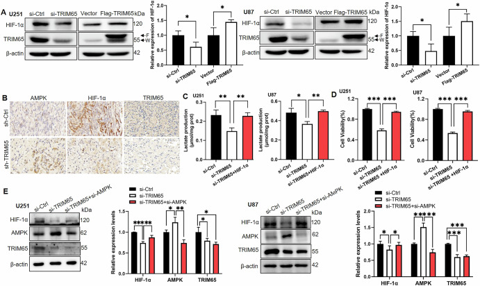
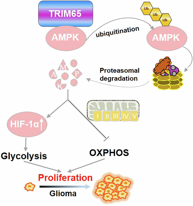
Glioma is the most common primary malignant brain tumor with high mortality and poor prognosis. Aerobic glycolysis is crucial for the malignant behavior of glioma by promoting their growth. Tripartite motif containing 65 (TRIM65) as an E3 ubiquitin ligase has been implicated in tumor progression, but its role and regulatory mechanism on aerobic glycolysis in glioma remains unclear. Here, it was demonstrated that TRIM65 was highly expressed in human glioma tissues and associated with poor prognosis. Moreover, TRIM65 knockdown inhibited the glioma cells proliferation in vitro and in vivo. RNA sequencing and biological verifications were performed to elucidate a novel mechanism underlying TRIM65 silencing attenuated glycolysis and enhanced OXPHOX to suppress the growth of glioma cells. Subsequently, we found that TRIM65 interacted with AMPK, a metabolic sensor, and mediated its K48-linkage ubiquitination and degradation though proteasomal pathway, thereby regulating HIF-1α-induced glycolysis. Importantly, the inhibitory effect of TRIM65 silencing on glycolysis was abrogated by AMPK knockdown or HIF-1α overexpression, indicating glucose metabolic reprogramming by TRIM65 is dependent on AMPK and HIF-1α pathway. These results reveal a new role for TRIM65/AMPK/HIF-1α axis in glioma cell proliferation and aerobic glycolysis, suggesting that TRIM65 may be a potential therapeutic target for intervention of glioma.


Conflict of interest statement

Competing interests: The authors declare no competing interests.

Figures

Fig. 1
Fig. 1. TRIM65 expression is upregulated in glioma and associated with poor survival of glioma patients.
A TRIM65 mRNA levels in normal tissues, LGG tissues and GBM tissues from TCGA and GTEx datasets. TPM transcripts per kilobase of exon model per million mapped reads. B TRIM65 protein levels in normal tissues and GBM tissues from CPTAC data. C IHC staining of a representative glioma tissue microarray with an anti-TRIM65 antibody. D TRIM65 expression level correlates positively with the grades of glioma. E Kaplan-Meier survival curve of 149 patients in the glioma tissue microarray based on TRIM65 expression. *p < 0.05; **p < 0.01, ***p < 0.001.
Fig. 2
Fig. 2. TRIM65 promotes glioma cell proliferation and tumor growth in vitro and in vivo.
A TRIM65 knockdown or overexpression efficiency in U251 and U87 cells was detected by Western blotting. F Flag-tagged TRIM65, W wild-type TRIM65. B TRIM65 was knocked down using TRIM65 siRNA or overexpressed using pCMV-Flag-TRIM65 plasmid in U251 and U87 cell lines. 24 h later, cells were seeded in 12-well plates. At indicated time, cells were counted. C Estimation of cell proliferation by EdU assay. Scale bar = 50 μm. D Estimation of cell proliferation by colony formation assay. E U87 cells were infected with sh-TRIM65 lentivirus (a titer of 1 × 108TU/mL) at a multiplicity of infection (MOI) of 10 for 48 h. Western blot assay for TRIM65 expression level in U87 cells stably expressing sh-Ctrl or sh-TRIM65. F U87 cells stably expressing sh-Ctrl or sh-TRIM65 were injected subcutaneously into the flanks of NOD-SCID mice. Two weeks later, tumors were dissected out. Representative images of the xenograft tumors in each group. G Tumor weight was measured. H Tumor volume was measured. I H&E staining of tumor and IHC staining using anti-Ki67 and anti-TRIM65 antibodies at the indicated treatment. Scale bar = 20 μm. Data are presented as mean ± SD. The experiments were independently repeated at least three times and the data of one representative experiment was shown. *p < 0.05; **p < 0.01, ***p < 0.001.
Fig. 3
Fig. 3. TRIM65 knockdown may be involved in glucose metabolic reprogramming of glioma.
A Volcano plot of DEGs between si-Ctrl and si-TRIM65 cells. B GSEA analysis. C GO analysis of the DEGs. The x-axis presents GO terms and y-axis presents the numbers of genes.
Fig. 4
Fig. 4. TRIM65 regulates aerobic glycolysis in glioma cells.
A The effect of TRIM65 knockdown on lactate production in U251 cells treated with or without oligomycin (Left panel). Lactate production was measured after TRIM65 overexpression in U251 cells (Right panel). B, C Western blot assay for HK1, PDK1 expression and OXPHOS in U251 cells knocked down (Left panel) or overexpressed TRIM65 (Right panel) using anti-HK1, anti-PDK1 and OXPHOS monoclonal antibody cocktail. F Flag-tagged TRIM65, W wild-type TRIM65. D Representative IHC images of xenograft tumors with anti-TRIM65, anti-PDK1 and anti-IDH3B. Scale bar = 20 μm. E Mitochondrial and glycolytic ATP were measured after TRIM65 knockdown in U251 cells. F MMP was detected by TMRE staining at the indicated treatment. Scale bar = 100 μm. G Cell viability was detected in U251 cells overexpressed TRIM65 in the presence or absence of 2-DG. Data are presented as mean ± SD. The experiments were independently repeated at least three times and the data of one representative experiment was shown. *p < 0.05; **p < 0.01, ***p < 0.001.
Fig. 5
Fig. 5. TRIM65 induces AMPK degradation through the K48-linkage ubiquitin-proteasome pathway.
The interaction between TRIM65 and AMPK was tested using anti-Flag (A) and anti-AMPK (B) antibodies for co-IP, followed by Western blot analysis. C Western blot analysis of AMPK protein expression in U251 cells with TRIM65 knockdown or overexpression. F Flag-tagged TRIM65, W wild-type TRIM65. D U251 cells were transfected with vector plasmid or Flag-TRIM65 and treated with 25 μg/mL CHX for different time points, followed by western blot analysis (Left panel). Related AMPK protein level was quantified at the indicated times (Right panel). E U251 cells were transfected with vector plasmid or Flag-TRIM65 in the presence or absence of MG132, followed by western blot analysis (Left panel). Related AMPK protein level was quantified (Right panel). FH U251 cells were co-transfected with Flag-TRIM65 and His-Ub in the presence or absence of MG132. Proteins were immunoprecipitated with AMPK antibody. The ubiquitination (F), K48-linkage ubiquitination (G) and K63-linkage ubiquitination (H) were detected. Poly-Ub chains were indicated. F Flag-tagged TRIM65, W wild-type TRIM65. Data are presented as mean ± SD. The experiments were independently repeated at least three times and the data of one representative experiment was shown. *p < 0.05; **p < 0.01, ***p < 0.001.
Fig. 6
Fig. 6. The attenuated glycolysis by TRIM65 silencing is abrogated by AMPK deletion in glioma cells.
A AMPK knockdown efficiency was detected by Western blotting. Cell viability (B), lactate production (C) and ATP content (D) were measured in U251 and U87 cells knocked down TRIM65 in the presence or absence of si-AMPK. E Western blot analysis of HK1, PDK1, AMPK and TRIM65 protein expression in U251 (Left panel) and U87 cells (Right panel) treated as indicated, and then the related protein level was quantified. Data are presented as mean ± SD. The experiments were independently repeated at least three times and the data of one representative experiment was shown. *p < 0.05; **p < 0.01, ***p < 0.001.
Fig. 7
Fig. 7. TRIM65 regulates the glycolysis process through AMPK and HIF-1α pathway.
A Western blot analysis of HIF-1α protein expression in U251 and U87 cells with TRIM65 knockdown or overexpression and then the related protein level was quantified. F Flag-tagged TRIM65, W wild-type TRIM65. B Representative IHC images of xenograft tumors with anti-TRIM65, anti-AMPK and anti-HIF-1α. Scale bar = 20 μm. Lactate production (C) and cell viability (D) were measured in U251 and U87 cells knocked down TRIM65 in the presence or absence of overexpressed HIF-1α. E Western blot analysis of HIF-1α protein expression in U251 and U87 cells treated as indicated. The related protein level was quantified. Data are presented as mean ± SD. The experiments were independently repeated at least three times and the data of one representative experiment was shown. *p < 0.05; **p < 0.01, ***p < 0.001.
Fig. 8
Fig. 8. Schematic diagram depicting the role and mechanism of TRIM65 in glioma.
TRIM65-catalized K48-linkage ubiquitylation and degradation of AMPK, which promotes HIF-1α-mediated aerobic glycolysis, as well as attenuating OXPHOS process, thus leading to the tumorigenesis of glioma.

Similar articles

References

    1. Weller, M. et al. Glioma. Nat. Rev. Dis. Prim.10, 33 (2024). - PubMed
    1. Louis, D. N. et al. The 2016 World Health Organization classification of tumors of the central nervous system: a summary. Acta Neuropathol.131, 803–820 (2016). - PubMed
    1. Stupp, R. et al. Radiotherapy plus concomitant and adjuvant temozolomide for glioblastoma. N. Engl. J. Med.352, 987–996 (2005). - PubMed
    1. Ghosh, D., Nandi, S. & Bhattacharjee, S. Combination therapy to checkmate Glioblastoma: clinical challenges and advances. Clin. Transl. Med.7, 33 (2018). - PMC - PubMed
    1. Hanahan, D. & Weinberg, R. A. Hallmarks of cancer: the next generation. Cell144, 646–674 (2011). - PubMed

LinkOut - more resources