. 2025 Jun 5;8(1):868.
doi: 10.1038/s42003-025-08299-y.

tRF16 affects NFKBIA stability and promotes osteoarthritis progression by regulating ALKBH5 expression in m6A-dependent manner

Affiliations

tRF16 affects NFKBIA stability and promotes osteoarthritis progression by regulating ALKBH5 expression in m6A-dependent manner

Chunhui Zhu et al. Commun Biol. .

Abstract

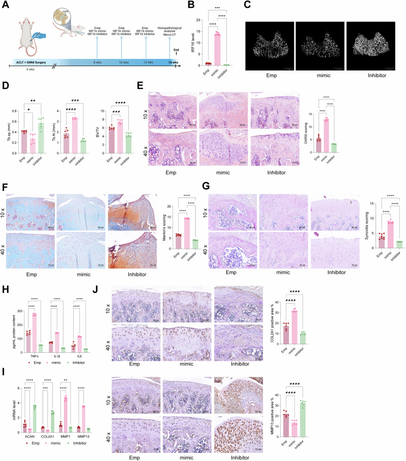
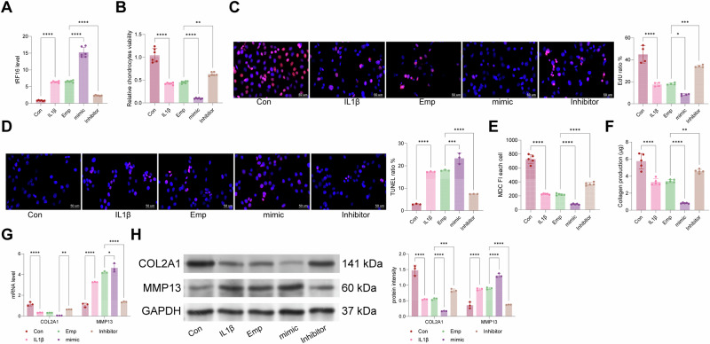
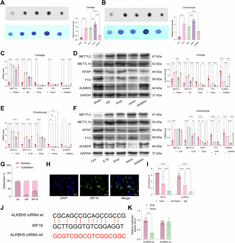
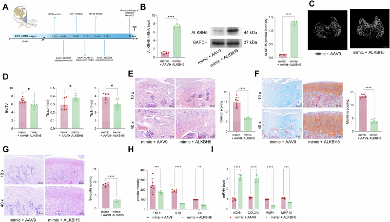
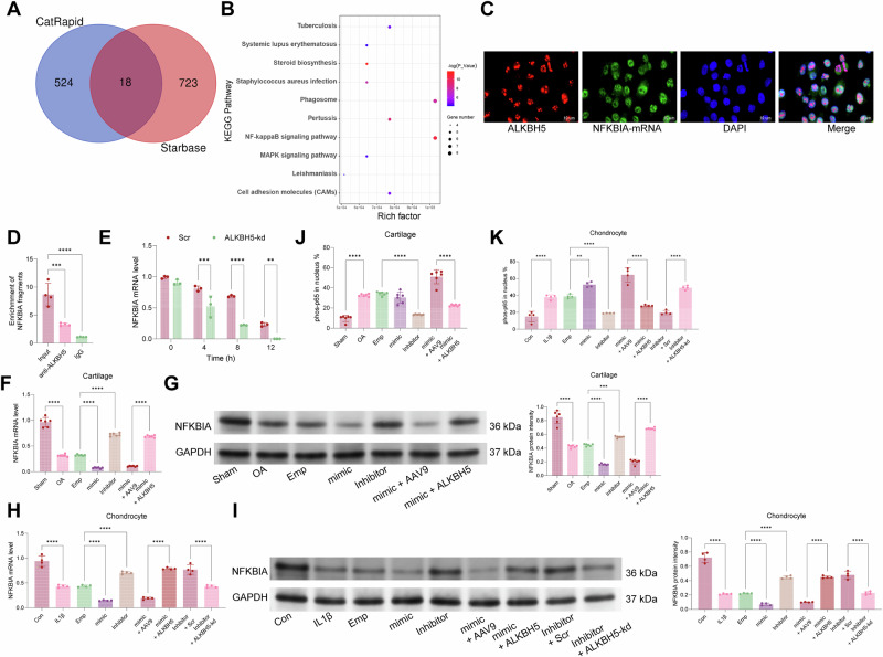
This study aimed to investigate explore the role of tRF16-ALKBH5 in osteoarthritis (OA). Differential expression of tRF in normal and OA tissues was analyzed using sequencing. OA rats model was established by destabilization of the medial meniscus and anterior cruciate ligament transaction surgeries. The tRF-16 inhibitor or mimic was injected into OA rats and OA symptoms were analyzed. We analyzed the m6A levels in tRF16 inhibitor-treated rat cartilage tissues, ALKBH5 levels, and the binding relationship between tRF16 and ALKBH5. The effect of ALKBH5 on OA rats was analyzed using specific antagonists. Chondrocytes were extracted to establish an OA cell model using IL-1β induction. The effects of tRF16, ALKBH5, and downstream genes on chondrocyte viability were verified. tRF-16 was overexpressed in OA patients and rat models. tRF16 inhibitor improved the symptoms of OA rats and inhibited autophagy and extracellular matrix degradation in IL-1β-induced chondrocytes. tRF16 reduced ALKBH5 expression by targeting ALKBH5, decreased NFKBIA mRNA stability, and activated the NF-kB pathway, thus exacerbating OA progression. Collectively, by binding to ALKBH5, tRF16 promotes the degradation of ALKBH5 and impairs the maintenance of NFKBIA mRNA stability by ALKBH5, promotes the nuclear translocation of phos-p65, leads to the secretion of inflammatory factors, exacerbates OA symptoms.


Conflict of interest statement

Competing interests: The authors declare no competing interests. Ethics approval and consent to participate: The studies involving human participants were reviewed and approved by the Ethics Committee and Institutional Review Board of the Third Affiliated Hospital of Nanjing Medical University ([2021]KY203-01). Written informed consent for participation in this study was provided by the participants. All animal experiments were approved by the Institutional Animal Care and Use Committee of the Third Affiliated Hospital of Nanjing Medical University.

Figures

Fig. 1
Fig. 1. tRF16 is significantly highly expressed in both OA patients and rats.
A, B Volcano and heat maps of smallRNAs differentially expressed in cartilage tissues of three normal subjects and three OA patients analyzed using Arraystar Human SmallRNA Expression Microarray; C flow diagram of a rat OA model established by meniscectomy. The illustration is created by Biorender.com; D microCT confirms arthritic symptoms in rats at 16 weeks postoperatively; E the Tb.N, Tb.sp and BV/TV value according to the micro-CT; FH, HE, PAS, and SAFG staining to detect pathological structural alterations in the cartilage tissues of rat knee joints; I RT-qPCR to detect tRF16 expression in the cartilage tissues of rats at 0, 4, 8, 12, and 16 weeks postoperatively; J RT-qPCR to detect the mRNA levels of Aggrecan, COL2A1, MMP1, and MMP13 in the cartilage tissues of rats; K the correlation between tRF16 expression in the knee joints of OA rats at week 16 and the Mankin score. Each group contained 6-8 rats; the data were presented as dot and whsikers and statistically analyzed using Student t-test or 2-way ANOVA, followed by Tukey’s multiple comparison test for post hoc tests, **P < 0.01, ***P < 0.001, ****P < 0.0001.
Fig. 2
Fig. 2. tRF16 inhibitor attenuates symptoms in OA rats.
A tRF16 mimic and tRF16 inhibitor were injected intracavitally into the knee joints of rats at the 8th, 12th, and 16th postoperative weeks. The illustration is created by Biorender.com; B RT-qPCR detected the expression level of tRF16 in the cartilage tissues of the rats at the 16th week; C microCT confirmed the arthritic symptoms of the rats at the 16th week postoperatively; D the Tb.N, Tb.sp and BV/TV value according to the micro-CT; EG, HE, PAS, and SAFG staining detected pathological structural changes in the cartilage tissues of rat knee joints; H ELISA to detect the levels of TNF-alpha, IL-1b, and IL-6 in the serum of rats at week 16; I RT-qPCR to detect the mRNA levels of Aggrecan, COL2A1, MMP1, and MMP13 in the cartilage tissues of rats. J IHC staining for MMP13 and COL2A1 in the cartilage tissues of rats. Each group contained 6 to 8 rats; the data were presented as dot and whiskers and statistically analyzed using 1-way or 2-way ANOVA, followed by post hoc test using Tukey’s multiple comparison test, **P < 0.01, ***P < 0.001, ****P < 0.0001.
Fig. 3
Fig. 3. tRF16 inhibitor alleviates ECM degradation in chondrocytes caused by IL-1β treatment.
A chondrocytes were transfected with constructed tRF16 inhibitor or mimic, and RT-qPCR verified the transfection efficiency; B, C, CCK-8 and EdU staining to detect chondrocyte viability and proliferative capacity; D, E, TUNEL staining and MDC staining to detect apoptosis and autophagy of chondrocytes; F the content of collagen produced by chondrocytes was detected; G, H RT-qPCR and WB detected the mRNA and protein levels of COL2A1 and MMP13 in chondrocytes. The experiments were repeated 3–5 times; the data were presented as dot and whiskers and statistically analyzed using 1-way or 2-way ANOVA, followed by Tukey’s multiple comparison test for post hoc testing, **P < 0.01, ***P < 0.001, ****P < 0.0001.
Fig. 4
Fig. 4. tRF16 binds to and causes degradation of ALKBH5.
A, B Dot blot assays to detect the levels of m6A RNA modification; C, D RT-qPCR and WB to detect the levels of METTL3/METTL14 and WTAP/FTO/ALKBH5 in rat cartilage tissue; E, F RT-qPCR and WB to detect the levels of m6A transferases METTL3/METTL14 and demethylases WTAP/FTO/ALKBH5 in chondrocytes; G, H Nuclear and cytoplasmic fragmentation experiments and FISH to confirm the subcellular localization of tRF16 in chondrocytes; I immunoprecipitation in chondrocytes using anti-Ago2 antibody and RT-qPCR to detect the levels of tRF16 and ALKBH5 mRNA in the precipitates; J, K luciferase reporter assay to analyze the relationship between tRF16 and ALKBH5 mRNA 3′-UTR binding relationship. The experiments were repeated 3 to 5 times; the data are presented as dot and whiskers and statistically analyzed using 1-way or 2-way ANOVA, followed by post hoc test using Tukey’s multiple comparison test, **P < 0.01, ***P < 0.001, ****P < 0.0001.
Fig. 5
Fig. 5. ALKBH5 impairs the exacerbating effect of tRF16 mimic on OA symptoms.
A tRF16 mimic was injected into the joint cavity of OA rats at weeks 4/8/12, and AAV9-ALKBH5 was injected into rats at weeks 6/10/14. The illustration is created by Biorender.com. B RT-qPCR and WB to detect the mRNA and protein levels of ALKBH5 in the cartilage tissues of rats at week 16. C microCT confirmed OA symptoms in the rats at week 16 after surgery; D the Tb.N, Tb.sp and BV/TV value according to the micro-CT; EG HE, PAS, and SAFG staining to detect pathological structural changes in the cartilage tissues of rat knee joints; H ELISA to detect the levels of TNF-alpha, IL-1b, and IL-6 in the serum of rats at week 16; I RT-qPCR to detect the mRNA levels of Aggrecan, COL2A1, MMP1, MMP13, and MMP13 in the cartilage tissues of rats. Each group contained 6 to 8 rats; the data were presented as dot and whiskers and statistically analyzed using 1-way or 2-way ANOVA, followed by post hoc test using Tukey’s multiple comparison test, **P < 0.01, ***P < 0.001, ****P < 0.0001.
Fig. 6
Fig. 6. ALKBH5-shRNA reverses the effect of tRF16 inhibitor on ECM degradation in chondrocytes.
A, B tRF16 mimic-transfected cells were further transfected with ALKBH5, while tRF16 inhibitor-transfected cells were further transfected with ALKBH5-shRNA. RT-qPCR and WB were used to detect the mRNA and protein levels of ALKBH5 in the cells. C, D CCK-8 and EdU staining to detect the viability and proliferation of chondrocytes; E, F TUNEL staining and MDC staining to detect apoptosis and autophagy of chondrocytes; G the content of collagen secreted by chondrocytes was detected; H, I RT-qPCR and WB to detect the mRNA and protein levels of COL2A1 and MMP13 in chondrocytes. The experiments were repeated 3–5 times; the data were presented as dot and whiskers and statistically analyzed using 1-way or 2-way ANOVA, followed by post hoc test using Tukey’s multiple comparison test, **P < 0.01, ***P < 0.001, ****P < 0.0001.
Fig. 7
Fig. 7. ALKBH5 maintains NFKBIA mRNA stability through m6A.
A Starbase and CatRapid Omics predicted the mRNAs that can bind to ALKBH5; B KEGG enrichment analysis of 18 intersecting genes obtained from cross-screening; C, D fluorescence co-localization assay and RIP assay confirmed that ALKBH5 can bind to NFKBIA mRNA; E the stability of NFKBIA mRNA was detected after knockdown of ALKBH5 in HEK293T cells; F, G RT-qPCR and WB assay for the mRNA and protein levels of NFKBIA in rat cartilage tissues; H, I RT-qPCR and WB detection of NFKBIA mRNA and protein levels in chondrocytes; J, K immunofluorescence detection of phos-p65 nucleation ratio in rat cartilage tissue or chondrocytes. The experiments were repeated 3 to 5 times; the data were presented as dot and whiskers and statistically analyzed using 1-way or 2-way ANOVA, followed by post hoc test using Tukey’s multiple comparison test, **P < 0.01, ***P < 0.001, ****P < 0.0001.

Similar articles

References

    1. Martel-Pelletier, J. et al. Osteoarthritis. Nat. Rev. Dis. Prim.2, 16072 (2016). - PubMed
    1. Karsdal, M. A. et al. Disease-modifying treatments for osteoarthritis (DMOADs) of the knee and hip: lessons learned from failures and opportunities for the future. Osteoarthr. Cartil.24, 2013–2021 (2016). - PubMed
    1. Charlier, E. et al. Insights on molecular mechanisms of chondrocytes death in osteoarthritis. Int. J. Mol. Sci.17, 2146 (2016). - PMC - PubMed
    1. Li, B. et al. Pathological mechanism of chondrocytes and the surrounding environment during osteoarthritis of temporomandibular joint. J. Cell Mol. Med.25, 4902–4911 (2021). - PMC - PubMed
    1. Glyn-Jones, S. et al. Osteoarthritis. Lancet386, 376–387 (2015). - PubMed

LinkOut - more resources