

Searching for protein partners of short-chain 3-hydroxyacyl-CoA dehydrogenase (SCHAD) reveals keratin 8 as a novel candidate for interaction in pancreatic β-cells
- PMID: 40474078
- PMCID: PMC12139081
- DOI: 10.1186/s12860-025-00544-w
Searching for protein partners of short-chain 3-hydroxyacyl-CoA dehydrogenase (SCHAD) reveals keratin 8 as a novel candidate for interaction in pancreatic β-cells
Abstract
Background: Short-chain 3-hydroxyacyl-CoA dehydrogenase (SCHAD) is a ubiquitously expressed mitochondrial enzyme with a role in the degradation of fatty acids. Because the protein also is a negative regulator of insulin secretion in pancreatic β-cells, inactivating mutations in the SCHAD gene (HADH) cause congenital hyperinsulinism of infancy (CHI) and severe hypoglycemia. Here we sought to identify novel interaction partners of SCHAD that might be particularly relevant for the endocrine pancreas.
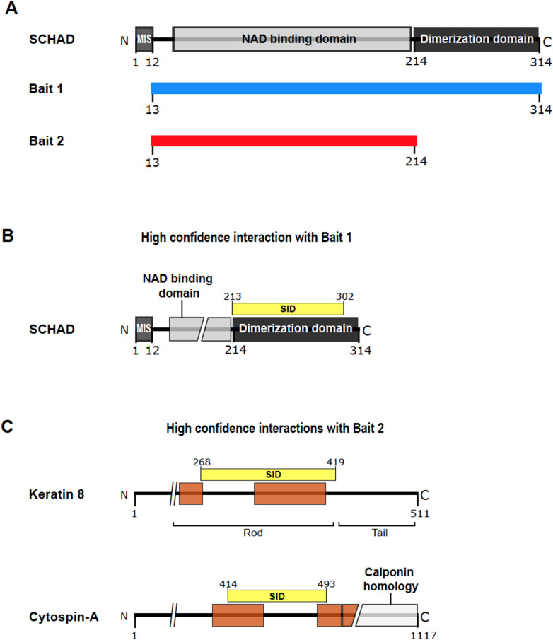
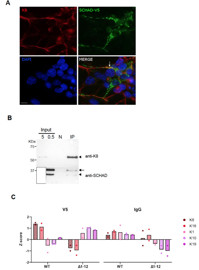
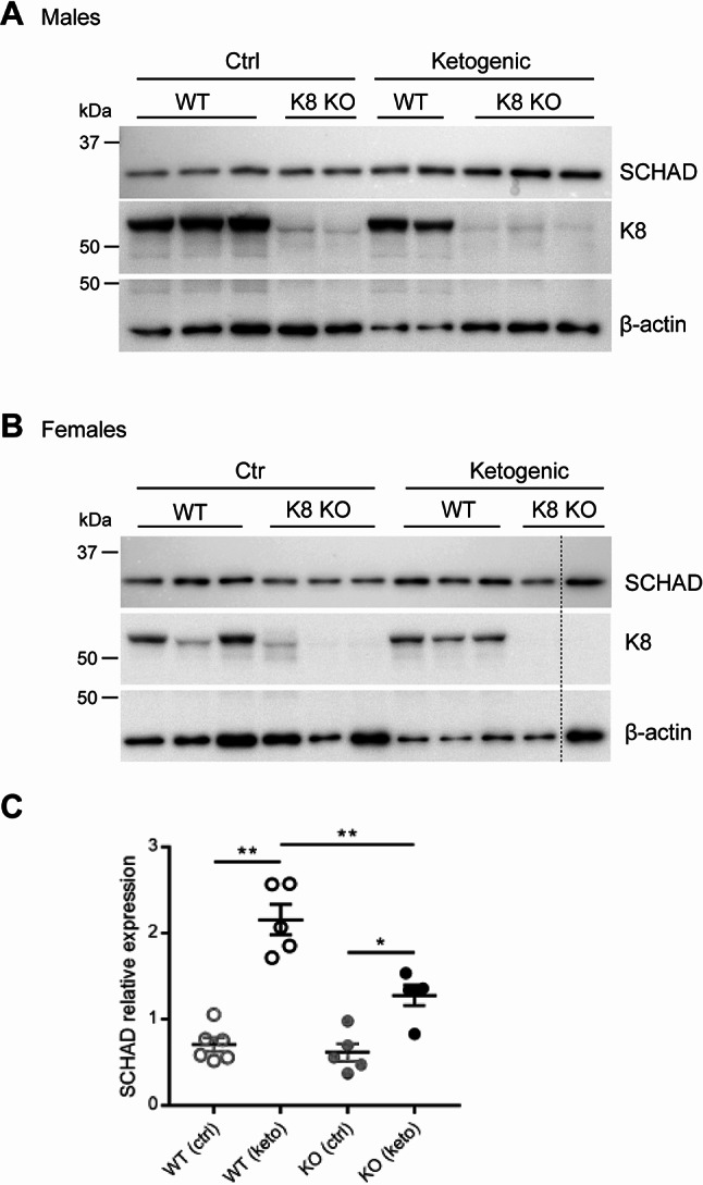
Results: Employing the SCHAD protein as bait, we performed yeast 2-hybrid screening of a cDNA library made from human islets of Langerhans. Surprisingly, the screening revealed the intermediate filament protein keratin 8 (K8) as a putative interaction partner of SCHAD with very high confidence. Previous reports have linked K8 to glucose homeostasis, and we confirmed the SCHAD interaction by co-immunoprecipitation in HEK293 cells. SCHAD and K8 expression were then characterized in the human β-cell model EndoC-βH1. By using proximity ligation assay, we demonstrated that stimulating the cells with a high level of glucose triggered a transient increase in the interaction. However, when studying knockout mice, we found that the loss of either K8 or SCHAD did not change the expression level of the other interaction partner. Still, when K8 knockout mice were challenged with a ketogenic diet, upregulation of SCHAD expression was blunted compared to the upregulation observed in wildtype littermates.
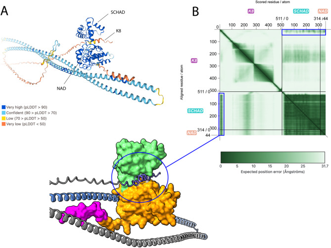
Conclusions: We propose that the SCHAD protein interacts with K8 in a way that might be relevant for proper functioning of the pancreatic β-cell. Whether the SCHAD-K8 interaction influences the phenotype of CHI remains to be demonstrated.
Keywords: HADH; KRT8; Congenital hyperinsulinism of infancy; Intermediate filaments; Keratin 8; Proximity ligation assay; SCHAD; Short-chain 3-hydroxyacyl-CoA dehydrogenase; Yeast 2-hybrid screening.
© 2025. The Author(s).
Conflict of interest statement
Declarations. Ethics approval and consent to participate: These animal studies were approved by the Institutiona Review Board of Joslin Diabetes Center and were in accordance with National Institutes of Heath (NIH) guidelines. Consent for publication: Not applicable. Competing interests: The authors declare no competing interests.
Figures


Similar articles
-
Mechanism of hyperinsulinism in short-chain 3-hydroxyacyl-CoA dehydrogenase deficiency involves activation of glutamate dehydrogenase.J Biol Chem. 2010 Oct 8;285(41):31806-18. doi: 10.1074/jbc.M110.123638. Epub 2010 Jul 29. J Biol Chem. 2010. PMID: 20670938 Free PMC article.
-
Functional evaluation of 16 SCHAD missense variants: Only amino acid substitutions causing congenital hyperinsulinism of infancy lead to loss-of-function phenotypes in vitro.J Inherit Metab Dis. 2021 Jan;44(1):240-252. doi: 10.1002/jimd.12309. Epub 2020 Sep 28. J Inherit Metab Dis. 2021. PMID: 32876354
-
Deficiency of the metabolic enzyme SCHAD in pancreatic β-cells promotes amino acid-sensitive hypoglycemia.J Biol Chem. 2023 Aug;299(8):104986. doi: 10.1016/j.jbc.2023.104986. Epub 2023 Jun 29. J Biol Chem. 2023. PMID: 37392854 Free PMC article.
-
Genetic pathogenesis, diagnosis, and treatment of short-chain 3-hydroxyacyl-coenzyme A dehydrogenase hyperinsulinism.Orphanet J Rare Dis. 2021 Nov 4;16(1):467. doi: 10.1186/s13023-021-02088-6. Orphanet J Rare Dis. 2021. PMID: 34736508 Free PMC article. Review.
-
3-Hydroxyacyl-CoA dehydrogenase and short chain 3-hydroxyacyl-CoA dehydrogenase in human health and disease.FEBS J. 2005 Oct;272(19):4874-83. doi: 10.1111/j.1742-4658.2005.04911.x. FEBS J. 2005. PMID: 16176262 Review.
References
-
- Velde CD, Reigstad H, Tjora E, Guthe HJT, Hansen EV, Molven A, Njølstad PR. Congenital hyperinsulinism. Tidsskr nor Laegeforen 2023: 143(18). - PubMed
-
- Maiorana A, Dionisi-Vici C. Hyperinsulinemic hypoglycemia: clinical, molecular and therapeutical novelties. J Inherit Metab Dis. 2017;40(4):531–42. - PubMed
-
- Clayton PT, Eaton S, Aynsley-Green A, Edginton M, Hussain K, Krywawych S, Datta V, Malingré HEM, Berger R, van den Berg IET. Hyperinsulinism in short-chain L-3-hydroxyacyl-CoA dehydrogenase deficiency reveals the importance of β-oxidation in insulin secretion. J Clin Invest. 2001;108(3):457–65. - PMC - PubMed
-
- Molven A, Matre GE, Duran M, Wanders RJ, Rishaug U, Njølstad PR, Jellum E, Søvik O. Familial hyperinsulinemic hypoglycemia caused by a defect in the SCHAD enzyme of mitochondrial fatty acid oxidation. Diabetes. 2004;53(1):221–7. - PubMed
MeSH terms
Substances
LinkOut - more resources
Full Text Sources
Research Materials
Miscellaneous
