Review
. 2025 Jun 6;13(1):32.
doi: 10.1186/s40560-025-00794-y.

Disseminated intravascular coagulation

Affiliations
Review

Disseminated intravascular coagulation

Satoshi Gando et al. J Intensive Care. .

Abstract

Background: Disseminated intravascular coagulation (DIC) is characterized by systemic coagulation activation, anticoagulation pathway impairment, and persistent fibrinolysis suppression, resulting in widespread microvascular thrombosis, followed by hemorrhagic consumption coagulopathy and multiple organ dysfunction syndrome. This article aimed to provide a comprehensive and updated DIC overview.

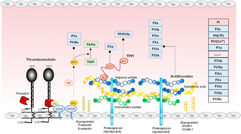
Main body: The International Society on Thrombosis and Hemostasis provides definitions, underlying disorders, diagnostic algorithms, and management guidelines for DIC. Two clinical features of DIC are hemorrhagic consumption coagulopathy, characterized by oozing and difficult-to-control bleeding, and microvascular thrombosis, leading to dysfunctions in multiple vital organs. Histones derived from cellular damage play central roles in the innate-immune-based coagulation model, comprising the initiation, amplification, propagation, and reinforcement phases, which, if dysregulated, develop into DIC. Thus, the innate immune-mediated pathogenic pathways in DIC have become clear. Cell death, damage-associated molecular patterns (including histones), crosstalk between hypoxic inflammation and coagulation, and the serine protease network (comprising coagulation and fibrinolysis, the Kallikrein-Kinin system, and complement pathways) play major roles in DIC pathogenesis. Conversely, these pathogenic pathways and DIC synergistically contribute to organ dysfunction, leading to poor prognoses. Effective DIC management requires treating the underlying condition, along with substitution therapies and, in some cases, antifibrinolytics. Anticoagulant use has been extensively debated; however, the selection of optimal target patients could optimize their application and improve patient outcomes in the near future.

Conclusions: This review provides an updated overview of DIC, aiming to help readers understand various aspects of DIC today.

Keywords: Cell death; Complement; Disseminated intravascular coagulation (DIC); Hemorrhage; Histone; Hypoxia; Inflammation; Innate immunity; Management; Organ dysfunction.


Conflict of interest statement

Declarations. Ethics approval and consent to participate: Not applicable. Consent for publication: Not applicable. Competing interests: The authors declare no competing interests.

Figures

Fig. 1
Fig. 1
Coagulation pathways and the Kallikrein–Kinin system. Injury-, PAMP-, or DAMP-mediated tissue factor exposure or expression in the circulation from tissue factor-bearing cells (figure assumes endothelial cells) triggers coagulation initiation, amplification, and propagation phases. In the innate immune-based coagulation model, histones play central roles in all three phases and reinforce coagulation by physiologically suppressing anticoagulation systems. Thrombin and tissue hypoxia under fibrin clots stimulate t-PA release from Weibel–Palade bodies in the endothelium, inducing fibrinolysis. Various DAMPs activate FXII, initiating KKS, where FXIIa, kallikrein, and t-PA-released Weibel–Palade bodies via bradykinin binding to KB2R convert plasminogen into plasmin. Bradykinin and its active metabolite DABAK induce inflammatory cytokine expression via KB2R and KB1R, respectively. Thus, coagulation, fibrinolysis, and inflammation are tightly coupled through the FXIIa and KKS axis, and serine proteases (*) have been well known to activate complement pathways, as depicted in Fig. 5. DABAK des-Arg9-bradykinin, DAMPs damage-associated molecular patterns, HMWK high-molecular weight kininogen, KB1R, KB2R bradykinin B1 and B2 receptors, KKS Kallikrein–Kinin system, NETs neutrophil extracellular traps, PAI-1 plasminogen activator inhibitor-1, PAMPs pathogen-associated molecular patterns, TF tissue factor, t-PA tissue-type plasminogen activator
Fig. 2
Fig. 2
Anticoagulation pathways and glycocalyx. Glycocalyx comprises glycoproteins, proteoglycans, and glycosaminoglycans. Thrombin binds to endothelial surface glycoprotein, thrombomodulin, via its epidermal growth factor-like domains 5 and 6. The formed complex activates protein C binding to EPCR to generate APC. The APC complexes with PS, degrading FVa and FVIIIa and accelerating thrombin-mediated TAFI activation to TAFIa, resulting in coagulation and fibrinolysis inhibition. TFPI comprises three Kunitz-type serine protease inhibitor domains (K1, K2, and K3), binding to glycosaminoglycan heparan sulfate through the K3 domain. K1 and K2 inhibit tissue factor/FVIIa complex and FXa, respectively. Proteoglycan syndecan has four subtypes. Syndecan 4 complexes with heparan sulfate and chondroitin sulfate, facilitating antithrombin binding to syndecan 4 heparan sulfate, thereby inhibiting factors IIa, VIIa, IXa, Xa, XIa, and XIIa. In summary, all coagulation factors except FI (fibrinogen), FIV (Ca2+), and FXIII (transglutaminase) can be controlled by the three anticoagulant systems. In addition, the anti-inflammatory properties of APC, TFPI, and antithrombin are well known. Glycoproteins such as selectins, ICAM-1, and VCAM-1 are upregulated or induced upon insults, mediating leucocyte rolling, tethering, adhesion, and transmigration across the endothelium. APC activated protein C, EPCR endothelial protein C receptor, ICAM-1 intercellular adhesion molecule-1, PC protein C, PS protein S, TAFI thrombin-activatable fibrinolysis inhibitor, TAFIa activated TAFI, TF tissue factor, TFPI tissue factor pathway inhibitor, VCAM-1 vascular cell adhesion molecule-1
Fig. 3
Fig. 3
Pyroptosis triggers the release of tissue factor-positive extracellular vesicles. PAMPs and DAMPs activate the NLRP3 inflammasome through the NLR family NLRP3. LPS complexed with HMGB1 translocate to cells through RAGE, resulting in caspase11 (noncanonical inflammasome) activation. Two signals are required to activate the NLRP3 inflammasome. Transcriptional expressions of pro-IL1β and pro-IL18 via NFκB activation by PAMP and DAMP recognition via TLRs or C5a/C5aR act as priming signals. The second signals are K+ efflux, Ca2+ influx, ROS release from dysfunctional mitochondria, and ATP influx that activate inflammasomes. Canonical and noncanonical inflammasomes with activated caspases 1 and 11, respectively, cleave GSDMD to produce GSDMD-N, forming GSDMD-N pores, which release IL1β, IL18, and small DAMPs. The pore allows Ca2+, Na+, and H2O influx and K+ efflux. The Ca2+ activates phospholipid scramblase, exposing phosphatidylserine to the outer membrane, which changes tissue factors from encrypted to decrypted forms. Water-induced cell ballooning and swelling are followed by NINJ1-dependent plasma membrane rupture, releasing tissue factor-positive extracellular vesicles, HMGB1, and large DAMPs. Cytosolic DNA sensor cGAS/cGAMP/STING-induced Ca2+ release from ER is triggered by mitDNA and nDNA from dysfunctional mitochondria or nuclei. The dysfunctional mitochondria also release ROS. Both Ca2+ and ROS activate inflammasome caspases. ATP, adenosine triphosphate; ATP2A2, ATPase sarcoplasmic/endoplasmic reticulum Ca2+ transporting protein 2; CARD caspase activation recruitment domain, cGAMP cyclic GMP–AMP, cGAS cyclic GMP–AMP synthase, DAMPs damage-associated molecular patterns, ER endoplasmic reticulum, GSDMD gasdermin D, HMGB1 high-mobility group box-1, ITPR1 inositol 1,4,5-trisphosphate receptor type 1, IL interleukin, LPS lipopolysaccharide, mitDNA mitochondria DNA, nDNA nuclear DNA, NFκB nuclear factor-κB, NLR NOD-like receptor, NLRP3 NLR pyrin domain-containing protein3, NOD nucleotide-binding oligomerization domain, NINJ1 nerve injury-induced protein 1, PAMPs pathogen-associated molecular patterns, RAGE receptor for advanced glycan end-products, ROS reactive oxygen species, STING stimulator of interferon gene, TF tissue factor, TLR toll-like receptor
Fig. 4
Fig. 4
Bidirectional interplays among hypoxia, inflammation, and coagulation. Normoxia: HIF-1α hydroxylation by PHD and FIH is facilitated by spare non-mitochondrial oxygen, leading to ubiquitination by VHL, which results in proteasomal degradation and transcriptional suppression by FIH, preventing HIF-1α binding to transcriptional co-activator protein p300/CBP in the nucleus. Hypoxia: absence of spare oxygen inhibits HIF-1α hydroxylation by PHD and FIH, resulting in its translocation to the nucleus to recruit HIF-1β and binding to p300/CBP and HRE. This transcriptional complex leads to the expression or downregulation of HIF-1α target genes, including inflammatory cytokines. One of the main pathogeneses of DIC, inflammatory cytokines, conversely induce HIF-1α expression. Tissue hypoxia by DIC enhances HIF-1α translocation to the nucleus. Therefore, bidirectional interplays exist among hypoxia, inflammation, and coagulation, playing pivotal roles in DIC pathogenesis. Ang angiopoietin, CBP CREB-binding protein, CREB cytoplasmic polyadenylation element binding protein, DIC disseminated intravascular coagulation, FIH factors inhibiting HIF, HIF hypoxia-inducible factor, HRE hypoxic responsive element, IL interleukin, PAI-1 plasminogen activator inhibitor 1, PHD prolyl hydroxylase, TFPI tissue factor pathway inhibitor, TNF tumor necrosis factor, Ub ubiquitin, VEGF vascular endothelial growth factor, VHL von Hippel–Lindau protein
Fig. 5
Fig. 5
Serine protease network and its inhibitory mechanisms. Activated coagulation factors, thrombin, plasmin, and kallikrein noncanonically activate complements, which in turn activate platelets, coagulation, and endothelial cells associated with fibrinolysis suppression by PAI-1 through C3/C3aR and C5/C5aR1. Anticoagulation pathways are also dampened by C4bBP binding to protein S and C5a-induced glycocalyx degradation and shedding, reducing activated protein C and antithrombin functions, respectively. MAC-injured plasma membrane releases tissue factor-bearing extracellular vesicles and increased intracellular Ca2+ exposes phosphatidylserine on the outer membrane by scramblase activation. These processes are controlled by C1-INH, antithrombin, TFPI, and TAFIa, as depicted in the figure. Furthermore, antithrombin indirectly controls complement pathways by inhibiting kallikrein, factors XIIa, XIa, Xa, and IXa, and thrombin. Dysregulation of these control mechanisms in severe insults can easily progress to DIC. C1-INH C1 esterase inhibitor, C4BP C4b binding protein, KKS Kallikrein–Kinin system, MAC membrane attack complex, MASP mannose-binding lectin-associated serine protease, TAFIa activated thrombin-activatable fibrinolysis inhibitor, TFPI tissue factor pathway inhibitor
Fig. 6
Fig. 6
Hazard of in-hospital mortality by DIC score and disease severity. The optimal target population for anticoagulant therapy in sepsis is not all patients with sepsis or sepsis-induced coagulopathy, but specifically those with an established DIC diagnosis and high disease severity. A The arrow indicates that anticoagulant therapy (blue plate) decreased the relative hazard ratio for in-hospital mortality compared with the control (non-anticoagulant; pink plate) in patients with sepsis diagnosed as having DIC and high APACHE II scores (shaded areas indicate 95% confidence interval). Reprint permission (Georg Thieme Verlag KG, No.5965741147872. Feb 11, 2025). B Anticoagulant therapy (blue plate) decreased hazard ratio of in-hospital mortality compared with the control (non-anticoagulant; red plate) in patients with higher JAAM DIC score and PTINR ≥ 1.5 in patients with sepsis. Reprint permission under a Creative Commons Attribution 4.0 International License. APACHE Acute Physiology and Chronic Health Evaluation, DIC disseminated intravascular coagulation, ISTH International Society on Thrombosis and Hemostasis, JAAM Japanese Association for Acute Medicine, PTINR prothrombin time international normalized ratio
Fig. 7
Fig. 7
Diverse disorders evoke innate immunity comprising local inflammation associated with immunothrombosis at the site of insults, which aims to maintain homeostasis, leading to recovery. When the insults are severe and persistent, dysregulated innate immunity cannot restrict the insults locally, developing into disseminated immunothrombosis with systemic inflammation, which is defined as DIC, which can lead to MODS and death of the patient. In addition to treating underlying DIC disorders, DIC treatments with systemic inflammation and organ dysfunction are mandatory to improve patient outcomes. DIC disseminated intravascular coagulation, MODS multiple organ dysfunction syndrome, SIRS systemic inflammatory response syndrome

References

    1. Gando S, Levi M, Toh CH. Disseminated intravascular coagulation. Nat Rev Dis Primers. 2016;2:16037. - PubMed
    1. McKay DG. Progress in disseminated intravascular coagulation. Calif Med. 1969;111:186–98. - PMC - PubMed
    1. McKay DG. Progress in disseminated intravascular coagulation. II Calif Med. 1969;111:279–90. - PMC - PubMed
    1. Colman RW, Robboy SJ, Minna JD. Disseminated intravascular coagulation (DIC): an approach. Am J Med. 1972;52:679–89. - PubMed
    1. Esmon CT, Fukudome K, Mather T, Bode W, Regan LM, Stearns-Kurosawa DJ, et al. Inflammation, sepsis, and coagulation. Haematologica. 1999;84:254–9. - PubMed

Grants and funding

LinkOut - more resources