. 2025 Jun 5;18(1):209.
doi: 10.1186/s13071-025-06854-4.

PD-1-dependent therapeutic effect of Trichinella spiralis cystatin on myocardial infarction in a mice model

Affiliations

PD-1-dependent therapeutic effect of Trichinella spiralis cystatin on myocardial infarction in a mice model

Weixiao Zhang et al. Parasit Vectors. .

Abstract

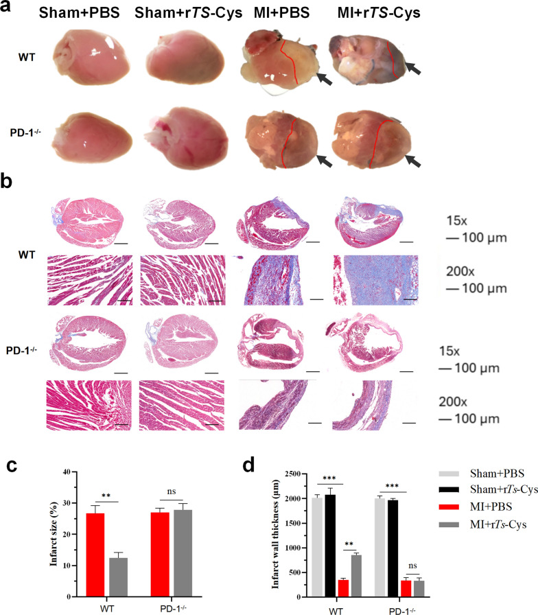
Background: Ischemia-induced inflammation is the primary pathological mechanism underlying cardiac tissue injury caused by myocardial infarction (MI). Trichinella spiralis cystatin (Ts-Cys) has been shown to regulate macrophage polarization and alleviate various inflammatory and immune-related diseases. Programmed cell death-1 (PD-1) is a crucial checkpoint receptor molecule and highly involved in maintaining immune tolerance. In this study, our aims were to investigate whether recombinant Ts-Cys protein (rTs-Cys) could be used to treat MI with recruited macrophage-dominant myocardial inflammation and whether PD-1 is involved in the immunomodulation of Ts-Cys in inflammatory diseases.

Methods: MI models were established in wild-type (WT) and PD-1 knockout (PD-1-/-) mice, followed by the intraperitoneal injection of rTs-Cys. The survival rates of mice were observed for 28 days post-surgery and treatment. Cardiac function was assessed by echocardiography. Histopathological evaluation of heart tissue affected by infarction was conducted to examine local inflammatory cell infiltration and cardiac tissue damage. Real-time quantitative PCR was used to detect messenger RNA expression levels of vascular endothelial growth factor (VEGF) and the macrophage surface markers inducible nitric oxide synthase and arginase-1 in the MI area. Serological levels of cytokines, including tumor necrosis factor-alpha (TNF-α), interleukin-6 (IL-6), interleukin-10 (IL-10) and transforming growth factor-beta (TGF-β), were measured using an enzyme-linked immunosorbent assay. Bone marrow-derived macrophages from WT and PD-1-/- mice were used to assess the effects of rTs-Cys on macrophage polarization in vitro.

Results: In WT mice, rTs-Cys treatment significantly improved the 28-day survival rate and cardiac function, reduced local inflammatory cell infiltration and cardiac pathological damage and increased VEGF expression levels of MI mice. The therapeutic effect of rTs-Cys was associated with macrophage polarization from the pro-inflammatory M1 phenotype to the regulatory M2 phenotype with reduced levels of inflammatory cytokines (TNF-α and IL-6) and increased levels of regulatory cytokines (IL-10 and TGF-β), as determined by both in vivo and in vitro tests. However, this therapeutic effect of rTs-Cys on MI was significantly reduced in PD-1-/- mice, as reflected by the higher level of M1 macrophages, elevated levels of inflammatory cytokines and decreased levels of regulatory cytokines.

Conclusions: rTs-Cys promotes M2-type polarization of macrophages through the PD-1 pathway to alleviate MI in mice and is, therefore, a potential drug for the treatment of MI and other inflammation-related diseases with involvement of the PD-1 checkpoint molecule.

Keywords: Trichinella spiralis; Immunomodulation; Inflammation; Macrophage; Myocardial infarction; PD-1.


Conflict of interest statement

Declarations. Ethics approval and consent to participate: All animal experimental procedures are complied with the ethical guidelines of Bengbu Medical University and were approved by the Ethics Committee (Approval No. [2023]587). Consent for publication: Not applicable. Competing interests: The authors declare no competing interests.

Figures

Fig. 1
Fig. 1
Identification of knockout of the programmed death-1 (PD-1) in PD-1−/− mice by PCR with PD-1-specific primers. Lanes: bp, size of DNA ladder; 0, blank control; 1–9, mouse number. PD-1−/−, programmed PD-1 knockout mice; WT, wild-type mice
Fig. 2
Fig. 2
Treatment with rTs-Cys significantly improved the survival rate of MI mice with PD-1 expression. The 28-day survival rate of each group of mice after surgery and treatment (n = 20) is shown. Asterisks indicate a statistically significant difference in survival rate between groups at *P < 0.05 and **P < 0.01; ns, not significant. MI, Myocardial infarction; PBS, phosphate-buffered saline; PD-1−/−, programmed death-1 (PD-1) knockout mice; rTs-Cys, recombinant Trichinella spiralis cystatin; WT, wild-type mice
Fig. 3
Fig. 3
Treatment with rTs-Cys significantly improved the SV, LVEF and LVFS in MI mice with PD-1 expression. a Typical B-mode ultrasound image in mice from different groups. b Typical M-mode ultrasound image in mice from different groups. c Cardiac function parameters in different groups (n = 6). The results are presented as the mean ± standard error of the mean (SEM). Asterisks indicate a statistically significant difference in the respective parameter at *P < 0.05, **P < 0.01 and ***P < 0.001; ns, not significant. LVEF, Left ventricular ejection fraction; LVFS, left ventricular fractional shortening; MI, myocardial infarction; PBS, phosphate-buffered saline; PD-1−/−, programmed death-1 (PD-1) knockout mice; rTs-Cys, recombinant Trichinella spiralis cystatin; SV, stroke volume; WT, wild-type mice
Fig. 4
Fig. 4
Treatment with rTs-Cys significantly reduced histopathological damage in the hearts of MI mice with PD-1 expression. a Representative shape of the heart from the different groups of mice. b Representative Masson staining sections of infarcted heart tissues from the different groups of mice (magnification 15× and 200×). c Percentage of left ventricular infarction area. d Left ventricular infarct wall thickness. The results are presented as the mean ± standard error of the mean (SEM) (n = 6). Asterisks indicate a statistically significant difference between groups at **P < 0.01 and ***P < 0.001; ns, not significant. MI, Myocardial infarction; PBS, phosphate-buffered saline; PD-1−/−,programmed death-1 (PD-1) knockout mice; rTs-Cys, recombinant Trichinella spiralis cystatin; WT, wild-type mice
Fig. 5
Fig. 5
Hematoxylin/eosin (HE) staining of representative heart tissue sections within the infarcted area of the heart from the different groups of mice 7 days after the MI operation and treatment (magnification 200×, scale bar: 100 μm). MI, Myocardial infarction; PBS, phosphate-buffered saline; PD-1−/−, programmed death-1 (PD-1) knockout mice; rTs-Cys, recombinant Trichinella spiralis cystatin; WT, wild-type mice
Fig. 6
Fig. 6
Transcriptional levels of vascular endothelial growth factor (VEGF) in the MI area of hearts from the different groups of mice relative to glyceraldehyde 3-phosphate dehydrogenase (GADPH) measured by real-time quantitative PCR. The results are presented as the mean ± standard error of the mean (SEM) (n = 6). Asterisks indicate a statistically significant difference at **P < 0.01; ns, not significant. MI, Myocardial infarction; mRNA, messenger RNA; PBS, phosphate-buffered saline; PD-1−/−, programmed death-1 (PD-1) knockout mice; rTs-Cys, recombinant Trichinella spiralis cystatin; VEGF, vascular endothelial growth factor; WT, wild-type mice
Fig. 7
Fig. 7
rTs-Cys regulated inflammatory-related cytokine expression. a Serological levels of the pro-inflammatory cytokines TNF-α and IL-6 and regulatory cytokines IL-10 and TGF-β, as measured by quantitative enzyme-linked immunosorbent assay. b The messenger RNA transcriptional levels of TNF-α, IL-6, IL-10 and TGF-β relative to glyceraldehyde 3-phosphate dehydrogenase (GADPH) in infarcted heart tissue, as measured by real-time quantitative PCR. The results are presented as the mean ± standard error of the mean (SEM) (n = 6). Asterisks indicate a statistically significant difference at *P < 0.05 and ***P < 0.001; ns, not significant. IL, Interleukin; MI, myocardial infarction; PBS, phosphate-buffered saline; PD-1−/−, programmed death-1 (PD-1) knockout mice; rTs-Cys, recombinant Trichinella spiralis cystatin; VEGF, vascular endothelial growth factor; WT, wild-type mice
Fig. 8
Fig. 8
rTs-Cys-induced PD-1-dependent M2 macrophage polarization in infarcted heart tissues. a Relative mRNA expression level of iNOS and Arg-1 in the infarcted cardiac tissues of WT and PD-1−/− mice. The results are presented as the mean ± standard error of the mean (SEM) (n = 6). Asterisks indicate a statistically significant difference at *P < 0.05, **P < 0.01 and ***P < 0.001; ns, not significant. b Representative immunofluorescence staining of cardiac tissue sections from the MI zone in the different groups at 7 days post-surgery and treatment. Scale bars: 1×, 1000 μm; 10×, 100 μm. Arg-1, Arginase-1; iNOS, inducible nitric oxide synthase; MI, myocardial infarction; mRNA, messenger RNA; PBS, phosphate-buffered saline; PD-1−/−, programmed death-1 (PD-1) knockout mice; rTs-Cys, recombinant Trichinella spiralis cystatin; VEGF, vascular endothelial growth factor; WT, wild-type mice
Fig. 9
Fig. 9
Levels of inflammatory cytokines (TNF-α and IL-6) and regulatory cytokines (IL-10 and TGF-β) in the supernatant of BMDMs derived from WT and PD-1−/− mice co-incubated with LPS with or without rTs-Cys, as measured by quantitative enzyme-linked immunosorbent assay. The results are presented as the mean ± standard error of the mean (SEM). Asterisks indicate a statistically significant difference at *P < 0.05, **P < 0.01 and ***P < 0.001; ns, not significant. BMDMs, Bone marrow-derived macrophages, IL, interleukin; LPS, lipopolysaccharide; MI, myocardial infarction; PBS, phosphate-buffered saline; PD-1−/−, programmed death-1 (PD-1) knockout mice; rTs-Cys, recombinant Trichinella spiralis cystatin; TGF-β, transforming growth factor-beta; TNF-α, tumor necrosis factor-alpha; WT, wild-type mice
Fig. 10
Fig. 10
Flow cytometry was performed to detect CD86 (M1 phenotype) and CD206 (M2 phenotype) glycoproteins on BMDMs derived from both WT and PD-1−/− mice, co-incubated with LPS and rTs-Cys. The results are presented as the mean ± standard error of the mean (SEM) (n = 3). Asterisks indicate a statistically significant difference at *P < 0.05, **P < 0.01 and ***P < 0.001; ns, not significant. BMDMs, Bone marrow-derived macrophages; FITC, fluorescein isothiocyanate; FSC, SSC, forward and side scatter, respectively; LPS, lipopolysaccharide; PBS, phosphate-buffered saline; PD-1−/−, programmed death-1 (PD-1) knockout mice; rTs-Cys, recombinant Trichinella spiralis cystatin; WT, wild-type mice
Fig. 11
Fig. 11
rTs-Cys promotes M2-type polarization of macrophages through the PD-1 pathway to improve MI in mice. a Intraperitoneal (i.P.) injection of rTs-Cys in MI mice effectively alleviates the inflammatory response in the damaged heart and mitigates MI symptoms. b rTs-Cys regulates the release of inflammatory cytokines from macrophages, thereby improving the inflammatory microenvironment of the heart, reducing myocardial fibrosis, and enhancing VEGF expression levels. This regulation is PD-1 dependent, C rTs-Cys improves cardiac function and tissue damage in MI mice by promoting the polarization of macrophages from the M1 to M2 phenotype through PD-1 regulation. α-SMAL, Alpha-smooth muscle actin; IL, interleukin; PD-1, programmed death-1; LVEF, left ventricular ejection fraction; LVFS, left ventricular fractional shortening; rTs-Cys, recombinant Trichinella spiralis cystatin; SV, stroke volume; TGF-β, transforming growth factor-beta; TNF-α, tumor necrosis factor-alpha; VEGF, vascular endothelial growth factor; WT, wild-type mice

Similar articles

References

    1. Murphy A, Goldberg S. Mechanical complications of myocardial infarction. Am J Med. 2022;135:1401–9. - PubMed
    1. Lindahl B, Mills NL. A new clinical classification of acute myocardial infarction. Nat Med. 2023;29:2200–5. - PubMed
    1. Bajaj A, Sethi A, Rathor P, et al. Acute complications of myocardial infarction in the current era: diagnosis and management. J Investig Med. 2015;63:844–55. - PubMed
    1. Tsao CW, Aday AW, Almarzooq ZI, et al. Heart disease and stroke statistics—2023 update: a report from the American Heart Association. Circulation. 2023;147:e93–621. - PMC - PubMed
    1. Krittanawong C, Khawaja M, Tamis-Holland JE, et al. Acute myocardial infarction: etiologies and mimickers in young patients. J Am Heart Assoc. 2023;12:e029971. - PMC - PubMed