. 2025:3:100967.
doi: 10.37349/eaa.2025.100967. Epub 2024 Dec 17.

The therapeutic potential of glucagon-like peptide-1 receptor analogs for neuroinflammation in the setting of asthma

Affiliations

The therapeutic potential of glucagon-like peptide-1 receptor analogs for neuroinflammation in the setting of asthma

Courtney Lehman et al. Explor Asthma Allergy. 2025.

Abstract

Glucagon-like peptide-1 (GLP-1) is a hormone that regulates blood glucose levels and is produced by the enteroendocrine glands in the large and small intestines in response to the consumption of foods that contain carbohydrates, fats, and proteins. When GLP-1 is secreted, it acts on the pancreas to increase insulin production and secretion, while decreasing pancreatic glucagon secretion in order to lower serum glucose. However, GLP-1 also regulates metabolism through the gut-brain axis. While GLP-1 is primarily produced in the gut and released into the bloodstream, small quantities of it can also be synthesized in distinct areas of neurons located in the hindbrain. Recent studies have proposed that GLP-1 receptor (GLP-1R) agonists (GLP-1RAs) may protect against neuroinflammatory diseases. GLP-1RAs may also be a therapeutic target for asthma as animal models show that these drugs reduce allergen-induced airway inflammation, as the GLP-1R is expressed on lung epithelial and endothelial cells. There is a notable association between insulin resistance and the onset of asthma, particularly among obese people, with this association suggesting that metabolic dysfunction may play a role in asthma development. There is also evidence that there may be a link between asthma pathobiology and neuroinflammation, suggesting that GLP-1 and its analogs may regulate neuroinflammatory pathways that contribute to asthma pathogenesis. Interest is growing, though research remains limited, in how inflammation in the nervous system and lung might be linked. This review will explore how GLP-1R signaling could inhibit interdependent inflammation in both the lung and nervous system. This review will first focus on the inflammation that is known to exist in asthma, then pivot to the current state of neural regulation of asthma, and finally speculate on how GLP-1RA signaling could inhibit both neural and lung inflammation in asthma treatment.

Keywords: Glucagon-like peptide-1 receptor; asthma; glucagon-like peptide-1 receptor agonist; neuroinflammation.


Conflict of interest statement

Conflicts of interest The authors declare that they have no conflicts of interest.

Figures

Figure 1.
Figure 1.. Neuroimaging of white matter microstructure demonstrates differences between individuals with asthma and healthy controls.
(A) and (B) display sagittal and axial views of the brain, highlighting regions with significant group differences using a yellow-orange color scale. (C) presents the distribution of mean diffusivity (MD) values for each group, averaged over brain regions where MD was significantly higher in the asthma group. This visualization emphasizes the structural brain differences associated with asthma compared to controls Note. Reprinted with permission from “Neuroimaging and biomarker evidence of neurodegeneration in asthma” by Rosenkranz MA, Dean DC 3rd, Bendlin BB, Jarjour NN, Esnault S, Zetterberg H, et al. J Allergy Clin Immunol. 2022;149:589–98.e6 (https://doi.org/10.1016/j.jaci.2021.09.010). © 2021 American Academy of Allergy, Asthma & Immunology.
Figure 2.
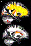
Figure 2.. Higher asthma severity reveals weaker white matter integrity.
(A) and (B) show sagittal (left) and axial (right) views that illustrate where asthma severity is significantly correlated with white matter microstructure changes. Regions in red highlight areas associated with overall changes in white matter microstructure across all diffusion-weighted imaging (DWI) metrics, while regions in blue indicate areas with increased mean diffusivity (MD) values. In panel A, affected regions include the inferior fronto-occipital fasciculus, superior longitudinal fasciculus, and uncinate fasciculus. In panel B, the highlighted regions include fibers in the superior longitudinal fasciculus, anterior thalamic radiation, and uncinate fasciculus Note. Reprinted with permission from “Neuroimaging and biomarker evidence of neurodegeneration in asthma” by Rosenkranz MA, Dean DC 3rd, Bendlin BB, Jarjour NN, Esnault S, Zetterberg H, et al. J Allergy Clin Immunol. 2022;149:589–98.e6 (https://doi.org/10.1016/j.jaci.2021.09.010). © 2021 American Academy of Allergy, Asthma & Immunology.
Figure 3.
Figure 3.. Elevated plasma biomarker levels are associated with white matter quality.
(A) and (B) show a sagittal view showing regions where plasma biomarker concentrations negatively correlate with neurite density index (NDI) values. In panel A, the yellow areas show the glial fibrillary acidic protein (GFAP) concentration that demonstrates a significant negative association with NDI, indicating neuroinflammation. In panel B, the red areas show the neurofilament light chain (NfL) concentration that reveals a similar negative relationship with NDI, indicating neurodegeneration. The affected regions primarily include fibers within the corona radiata and internal capsule, such as the corticospinal tract and thalamic radiations Note. Reprinted with permission from “Neuroimaging and biomarker evidence of neurodegeneration in asthma” by Rosenkranz MA, Dean DC 3rd, Bendlin BB, Jarjour NN, Esnault S, Zetterberg H, et al. J Allergy Clin Immunol. 2022;149:589–98.e6 (https://doi.org/10.1016/j.jaci.2021.09.010). © 2021 American Academy of Allergy, Asthma & Immunology.
Figure 4.
Figure 4.. A T-maze test was performed to assess memory acquisition and retention function in senescence-accelerated mouse prone 8 (SAMP8) mice.
(A) shows memory acquisition, measured as the number of trials required to make one active avoidance. (B) portrays memory retention, evaluated one week later by the number of trials needed to achieve five action avoidances in six consecutive trials. Vehicle-treated 10-month-old SAMP8 mice exhibited impaired memory compared to controls, including four-month-old untreated SAMP8 mice and 50% back crossed vehicle-treated SAMP8 mice. Notably, long-term liraglutide treatment restored memory retention in an older sample of eight mice two levels comparable to younger controls. **: P < 0.01; ***: P < 0.001 Note. Reprinted from “The GLP-1 Receptor Agonist Liraglutide Improves Memory Function and Increases Hippocampal CA1 Neuronal Numbers in a Senescence-Accelerated Mouse Model of Alzheimer’s Disease” by Hansen HH, Fabricius K, Barkholt P, Niehoff ML, Morley JE, Jelsing J, et al. J Alzheimers Dis. 2015;46:877–88 (https://doi.org/10.3233/JAD-143090). CC BY-NC.
Figure 5.
Figure 5.. The Glucagon-like peptide-1 receptor (GLP-1R) is expressed in human bronchi.
(A) displays a bar graph quantifying normalized GLP-1R immunoreactivity in medium and small bronchi, with pancreatic tissue serving as a positive control and untreated tissue as a negative control. (B) presents raw immunostaining data for GLP-1R in a bronchial section. (C) shows an interactive three-dimensional surface plot, highlighting GLP-1R expression. Results are based on samples from five different subjects. ASM: airway smooth muscle; ND: not detectable; **: P < 0.01; ***: P < 0.001 Note. Reprinted with permission from “Glucagon-Like Peptide 1 Receptor: A Novel Pharmacological Target for Treating Human Bronchial Hyperresponsiveness” by Rogliani P, Calzetta L, Capuani B, Facciolo F, Cazzola M, Lauro D, et al. Am J Respir Cell Mol Biol. 2016;55:804–14 (https://doi.org/10.1165/rcmb.2015-0311OC). © 2016 by the American Thoracic Society.
Figure 6.
Figure 6.. A schematic of the overall potential glucagon-like peptide-1 receptor agonist (GLP-1RA) indications and its connection to neuroinflammation and asthma.
BBB: blood-brain barrier

Similar articles

References

    1. Kanaya A, Yang M, Emala C, Mikami M. Chronic allergic lung inflammation negatively influences neurobehavioral outcomes in mice. J Neuroinflammation. 2022;19:210. - PMC - PubMed
    1. Slejko JF, Ghushchyan VH, Sucher B, Globe DR, Lin SL, Globe G, et al. Asthma control in the United States, 2008–2010: indicators of poor asthma control. J Allergy Clin Immunol. 2014;133:1579–87. - PubMed
    1. Papi A, Brightling C, Pedersen SE, Reddel HK. Asthma. Lancet. 2018;391:783–800. - PubMed
    1. Wu AY, Peebles RS. The GLP-1 receptor in airway inflammation in asthma: a promising novel target? Expert Rev Clin Immunol. 2021;17:1053–7. - PMC - PubMed
    1. Backman H, Räisänen P, Hedman L, Stridsman C, Andersson M, Lindberg A, et al. Increased prevalence of allergic asthma from 1996 to 2006 and further to 2016—results from three population surveys. Clin Exp Allergy. 2017;47:1426–35. - PubMed

LinkOut - more resources