

Ileosigmoid knot: A case report of the lethal twist
- PMID: 40475047
- PMCID: PMC12139662
- DOI: 10.1016/j.radcr.2025.04.069
Ileosigmoid knot: A case report of the lethal twist
Abstract
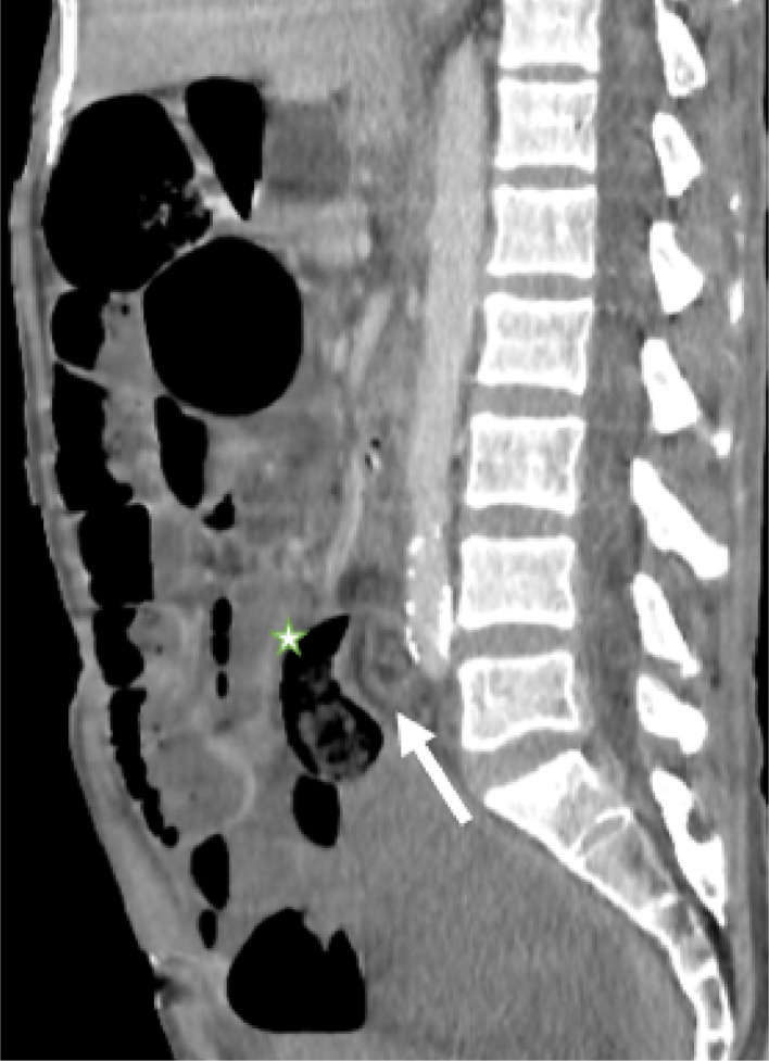
Ileosigmoid knot (ISK) is a rare and rapidly fatal surgical emergency characterized by the twisting of the ileum around the sigmoid colon, leading to acute bowel obstruction, ischemia, and gangrene. Due to its nonspecific clinical presentation, ISK is often misdiagnosed causing a delay of definitive treatment. The condition is most frequently reported in regions with high-fiber diets and anatomical predispositions, but remains a globally uncommon entity. Radiological imaging, particularly CT scans, plays a crucial role in preoperative diagnosis, with the whirl sign including sigmoid and ileum being a key diagnostic clue. However, most cases are only confirmed intraoperatively, and outcomes depend on the extent of bowel necrosis and the timeliness of surgical intervention. Mortality rates remain high, particularly in cases complicated by sepsis and multiorgan failure. We present the case of a 49-year-old male who developed acute abdominal pain, with CT imaging and laparotomy confirming an ileosigmoid knot, ultimately resulting in a fatal outcome.
Keywords: Computed tomography; Emergency radiology; Ileo-sigmoid knot; Intestinal volvulus; Laparotomy.
© 2025 The Authors. Published by Elsevier Inc. on behalf of University of Washington.
Figures


Similar articles
-
Ileosigmoid knotting: A rare cause of acute abdomen with fatal outcome in a 60-year-old man: Case report from a resource-limited setting.Int J Surg Case Rep. 2025 Aug;133:111641. doi: 10.1016/j.ijscr.2025.111641. Epub 2025 Jul 9. Int J Surg Case Rep. 2025. PMID: 40644986 Free PMC article.
-
Appendicular band syndrome leading to small bowel obstruction: A case report of a rare complication of acute appendicitis.Int J Surg Case Rep. 2025 Jul;132:111452. doi: 10.1016/j.ijscr.2025.111452. Epub 2025 May 19. Int J Surg Case Rep. 2025. PMID: 40435562 Free PMC article.
-
Surveillance for Violent Deaths - National Violent Death Reporting System, 50 States, the District of Columbia, and Puerto Rico, 2022.MMWR Surveill Summ. 2025 Jun 12;74(5):1-42. doi: 10.15585/mmwr.ss7405a1. MMWR Surveill Summ. 2025. PMID: 40493548 Free PMC article.
-
Understanding Glycogen Storage Disease Type IX: A Systematic Review with Clinical Focus-Why It Is Not Benign and Requires Vigilance.Genes (Basel). 2025 May 15;16(5):584. doi: 10.3390/genes16050584. Genes (Basel). 2025. PMID: 40428406 Free PMC article. Review.
-
Assessing the comparative effects of interventions in COPD: a tutorial on network meta-analysis for clinicians.Respir Res. 2024 Dec 21;25(1):438. doi: 10.1186/s12931-024-03056-x. Respir Res. 2024. PMID: 39709425 Free PMC article. Review.
References
-
- Atamanalp S.S., Disci E., Peksoz R., Atamanalp R.S., Atamanalp C.T. Ileosigmoid knotting: a review of 923 cases. Pak J Med Sci. 2022;38(3Part-I):711. https://pmc.ncbi.nlm.nih.gov/articles/PMC9002437/ [Accessed April 4, 2025]. Available from. - PMC - PubMed
-
- Lee S.H., Park Y.H., Won Y.S. The ileosigmoid knot: CT findings. Am J Roentgenol. 2000;174(3):685–687. https://www.ajronline.org/doi/10.2214/ajr.174.3.1740685 [Accessed February 6, 2025]. Available from: - DOI - PubMed
Publication types
LinkOut - more resources
Full Text Sources
