Review
. 2025 May 11:51:83-137.
doi: 10.1016/j.bioactmat.2025.04.037. eCollection 2025 Sep.

Progress in visible-light-activated photocatalytic coatings to combat implant-related infections: From mechanistic to translational roadmap

Affiliations
Review

Progress in visible-light-activated photocatalytic coatings to combat implant-related infections: From mechanistic to translational roadmap

Bruna E Nagay et al. Bioact Mater. .

Abstract

Biomedical and dental implants have enhanced healthcare but concurrently increased the risk of infections. Innovations in smart biomaterials, especially those responding to light stimuli through photocatalytic mechanisms, are emerging as promising solutions for activating targeted antimicrobial responses. While extensive reviews have provided insight into photocatalysis and its medical and environmental applications, limited focus has been given to solutions specifically tailored for implant contexts. The recent introduction of photocatalysis in the implant field, particularly visible-light-triggered photocatalytic coatings, represents a versatile approach to managing infections. These coatings offer on-demand reactive oxygen species generation, delivering antibacterial effects against a range of pathogens. Hence, this comprehensive review aims to summarize the latest advancements in design principles, physicochemical modifications, and surface optimizations, along with novel research concepts towards the achievement of visible-light-triggered photocatalytic antibacterial activity. Moreover, through a systematic search, this review discusses the current state-of-the-art regarding the antimicrobial efficacy of these biomaterials and the key factors influencing their performance, including microorganism type, photocatalyst properties, light source and intensity, and exposure time. Finally, it provides an in-depth discussion of current challenges, future directions, and regulatory considerations targeting biofilm-related implant treatments, offering guidance for future clinical adoption of multifunctional photocatalytic coatings in implant therapy.

Keywords: Biofilms; Coatings; Implants; Light; Photocatalysis.


Conflict of interest statement

The authors declare that they have no known competing financial interests or personal relationships that could have appeared to influence the work reported in this paper.

Figures

Image 1
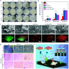
Graphical abstract
Fig. 1
Fig. 1
Schematic overview of the scope of the current review paper. This review focuses on the development of photocatalytic coatings to mitigate implant-related infections through light-activated mechanisms. These coatings depend on a light source with a wavelength compatible with the band gap of the semiconductor photocatalysts, facilitating the generation of reactive oxygen species (ROS) upon irradiation, which can damage bacterial components, ultimately leading to cell death. Emphasis is placed on engineering semiconductor materials to achieve visible-light photocatalytic activity, optimizing their physicochemical properties and performance. The review incorporates an evidence-based systematic search to summarize findings on the applicability of these coatings for (biomedical) implants. By leveraging interdisciplinary approaches from engineering, biomedical sciences, and chemistry, this work highlights the integration of material design, biological validation, and technological innovation, providing a foundation for advancing photocatalytically-active coatings into clinical practice.
Fig. 2
Fig. 2
Schematic representation illustrating the stages of biofilm formation on implant surfaces, key factors driving biofilm accumulation and pathogenesis, and antimicrobial strategies designed to overcome these processes. (a) The main phases of biofilm formation and spread include protein adsorption, microbial adhesion, biofilm development, maturation, and dispersal. Environmental conditions, such as reduced oxygen levels, availability of nutrients, metabolite accumulation, and mass transport mechanisms, can trigger microbial dysbiosis. This imbalance ultimately drives the transition from health to disease. (b) In parallel, several factors intrinsic to the material's surface influence microbial attachment and pathogenesis, further contributing to this shift. These surface-specific properties, including roughness, polarity charge, and electrochemical behavior, can create conditions conducive to bacterial colonization, thereby promoting adhesion and biofilm formation. Moreover, individual host responses, systemic conditions, and patient-specific habits can modulate the microbial profile and play a significant role in the progression of the pathogenesis process. (c) To address these challenges, antimicrobial surface mechanisms have been proposed, including contact-killing, antifouling, and releasing strategies. These mechanisms aim to either eliminate bacteria upon contact, prevent their adhesion, or release antimicrobial agents to inhibit microbial activity. (d) Among current antimicrobial strategies, surface modifications, antimicrobial coatings, superhydrophobic properties, and antibiotic-loaded surfaces have shown potential for inhibiting bacterial growth and preventing colonization on implant surfaces. By combining these approaches, it is possible to create more effective solutions to combat biofilm-related infections and improve implant longevity.
Fig. 3
Fig. 3
Schematic illustration of light-based approaches towards biofilm eradication. Photodynamic therapy (PDT), photothermal therapy (PTT), and photocatalysis each employ distinct antibacterial mechanisms but share the common principle of utilizing light to target and eliminate biofilms. PDT relies on a photosensitizer that, upon light activation, generates reactive oxygen species (ROS), leading to bacterial membrane damage and cell death. PTT leverages photothermal agents that absorb light and convert it into heat, raising the local temperature to induce bacterial destruction. This effect can be enhanced by plasmonic nanomaterials, which exhibit localized surface plasmon resonance, amplifying light absorption and heat generation. Photocatalysis, in turn, involves semiconductor materials that, upon light irradiation, generate electron-hole pairs, initiating oxidative reactions and ROS generation that kill bacteria. These techniques have been widely explored for medical and environmental applications, offering promising strategies for biofilm eradication.
Fig. 4
Fig. 4
Light-induced PTT effects for treating bacterial infections. (a-c) Copper-doped polyoxometalate nano-clusters (Cu-POM) exhibiting synergistic CDT and NIR-induced PTT effects. a) Schematic illustration of Cu-POM synthesis with bacterial cuproptosis-like death and macrophage activation functions. b) EPR spectra showing ·OH generation in H2O2solution, and c) metabolomic analysis using volcano plots, correlation analysis, and heat maps to depict differential gene expression. [134] Copyright 2023, Wiley. (d-g) Functionalized Ti implants with FeIIITA nanoparticles. d) Schematic representation of in vivo anti-infection evaluation, e) quantification of bacterial colony-forming units, f) schematic diagram illustrating coating synthesis, PTT effects under NIR irradiation, and macrophage-mediated immune responses, and g) real-time thermal imaging during NIR exposure. Copyright 2023, [135] Wiley.
Fig. 5
Fig. 5
Schematic illustration proposing the photocatalysis applications within the One Health concept. a) Photocatalysis leverages different light sources to address environmental, human, and animal health challenges. UV light and solar irradiation are widely applied in living environments, enabling self-cleaning surfaces, air pollutant removal, industrial wastewater treatment, and water purification, contributing to sustainable ecosystems. Visible light is used for medical applications, including antimicrobial therapies, infection control on implant surfaces, and wound healing, ensuring safe and effective treatment strategies while contributing to the prevention of antibiotic resistance. b) When focusing on human health applications, the type of light used becomes critical. UV light, while effective for photocatalysis, poses significant risks to tissues due to its carcinogenic and mutagenic effects, making it unsuitable for direct biomedical or implant dentistry use. Visible light, being safer and non-ionizing, emerges as the optimal choice for health-related photocatalytic applications, balancing efficacy and safety for patients, animals, and medical environments.
Fig. 6
Fig. 6
Photocatalytic coatings leverage ROS as their key antimicrobial agents, effectively killing bacteria through oxidative stress and structural disruption. Upon exposure to light with wavelengths matching the absorption spectrum of the semiconductor photocatalyst, ROS such as hydroxyl radicals, superoxide anions, and hydrogen peroxide are generated. These highly reactive species target and damage bacterial membranes, proteins, and nucleic acids, leading to the breakdown of essential cellular functions and ultimately causing bacterial inactivation.
Fig. 7
Fig. 7
Technological approaches for constructing photocatalytic coatings. Recent advancements in materials science and nanotechnology have revolutionized surface treatment and coating techniques, enabling the fabrication of efficient photocatalytic coatings with tailored properties. Key techniques include: a) sol-gel synthesis, a chemical process that involves hydrolysis and polycondensation reactions to produce metal oxide gels, allowing precise control of material composition and structure, making it ideal for fabricating thin, uniform coatings with customizable properties, a’) Spin-coating of a thin TiO2 film prepared by sol-gel chemistry. Reproduced with permission [221]. Copyright 2024, American Chemical Society. b) Anodization method, conducted in an electrolytic solution under constant voltage potential; this method promotes crystallization when submitted to annealing at high temperatures, enabling the growth of nanoscale coatings with enhanced photocatalytic and structural properties directly on substrates that can be easily decorated with nanoparticles using hydrothermal treatment. b’) Au-decorated nanoparticles on TiO2 nanotubes. Reproduced with permission [222]. Copyright 2020, MDPI. (c) Magnetron sputtering, a physical vapor deposition technique that bombards a target material with energetic particles at a low deposition rate, expelling atoms that condense into a thin, uniform film on a substrate, producing high-purity, adherent coatings with precise control over thickness and composition. c’) Ag nanoparticle-impregnated N-doped titania film. Reproduced with permission [223]. Copyright 2015, Springer Nature. d) Plasma electrolytic oxidation (PEO), also known as microarc oxidation (MAO), which employs electrical discharges in an electrolyte to generate porous, adherent crystalline coatings, making it especially suitable for biofunctional applications on complex metallic geometries. Its high surface area, hydrophilicity, and ability to incorporate chemical elements also enhance its effectiveness for photocatalytic coatings. d’) TiO2 nanoceramic coating. Reproduced with permission [185]. Copyright 2019, Elsevier.
Fig. 8
Fig. 8
Representative characterization of photocatalytic coatings highlighting the influence of coating thickness, wear resistance, corrosion, and tribocorrosion on photocatalytic performance and long-term durability. a) Cross-sectional FIB-TEM analysis of a photocatalytic plasma electrolytic oxidation coating showing tight interfacial anchorage to the titanium substrate, a dense nanocrystalline structure with anatase and rutile phases, and a ∼26 nm disordered outer layer. b) Scanning electron micrographs showing P. gingivalis biofilm formation and the wear effects of mechanical decontamination by curette and ultrasonic scaler. Reproduced with permission. [185] Copyright 2019, Elsevier. c) Wear and d,e) tribocorrosion scars as well as f) potentiodynamic polarization curves for the corrosion on uncoated and coated Ti6Al4V samples with TiO2/SrTiO3coating. Reproduced with permission [248] Copyright 2022, Elsevier. g) Schematic illustration of a visible-light-activated 2D g-C3N4/Cu2O/Cu coating exhibiting antibacterial photocatalytic activity and protection against microbial corrosion, with h) linear polarization resistance (LPR) images highlighting deeper corrosion on uncoated surfaces compared to coated ones (top to bottom). Reproduced with permission [249] Copyright 2024, Elsevier.
Fig. 9
Fig. 9
Strategies to extend the photocatalytic activity of semiconductors to the visible light region.a) Key strategies include metal doping, non-metal doping, co-doping, introducing oxygen vacancy, and forming heterojunctions. Examples of such approaches are: b) Doping of crystalline TiO2coating with metals, such as Bi. Reproduced with permission. [19] Copyright 2019, Elsevier. c) Nitrogen-doped TiO2coatings on reduced graphene oxide using sonochemical and hydrothermal methods, in which nitrogen doping was found to effectively reduce the band gap of TiO2, making it responsive to visible light. Reproduced with permission. [260] Copyright 2018, Elsevier. d) Introduction of oxygen vacancies to improve photocatalytic activity by reducing electron-hole pair recombination. Reproduced with permission. [261] Copyright 2019, Elsevier. e) The Cu2O/UiO-66/Bi2MoO6heterojunction enhances visible-light photocatalytic efficiency through improved charge separation and transfer via a double-Z scheme, with UiO-66 facilitating fast electron migration. Reproduced with permission. [262] Copyright 2025, Elsevier.
Fig. 10
Fig. 10
Representative example of a heterojunction- and oxygen-vacancy-based coating strategy responsive to visible–NIR light, illustrating its potential for biofilm-associated infection control and improved osseointegration. a) Schematic illustration of the TiO2/Bi2WO6 heterostructure in situ construction on titanium implant surfaces by first forming TiO2 nanowires via alkaline thermal treatment, followed by hydrothermal growth of Bi2WO6 nanocrystals. b) Plot of (αhv)12 versus photon energy (hv) for optical bandgap estimation. c) Insertion of TiO2/Bi2WO6-coated implant in a bone defect model with an induced bacterial infection. d) Images of the implantation sites and Giemsa staining showing bacterial localization around the implant surfaces. e) Spread plate images of E. coli and P. gingivalis cultured with different samples under dark conditions and NIR irradiation (808 nm, 1 W cm−2, 10 min in vitro). f) Micro-CT scans illustrating bone regeneration surrounding control and experimental implants after NIR irradiation. g) Schematic representation of ROS production by the TiO2/Bi2WO6 heterojunction and oxygen vacancies under NIR light exposure. h) Osteogenic differentiation was assessed through immunofluorescence and FE-SEM, revealing enhanced cell adhesion and morphology on TiO2/Bi2WO6 heterojunctions compared to control substrates [269]. Copyright 2024, Wiley.
Fig. 11
Fig. 11
Systematic search of relevant literature on antimicrobial effects of visible-light-triggered photocatalytic coatings for biomedical implant application. a) Schematic diagram of the systematic search methodology used. b) Publication timeline – Orange bars represent the number of studies focused on in vitro and in vivo analyses, while blue bars represent those concentrating on in vitro analyses. It is worth noting that studies conducting both in vitro and in vivo analyses have been counted twice to accurately reflect their contributions. c) Cake diagram representing the frequency of substrates used, in which darker colors represent more studies (Abbreviations – Ti: titanium, SS: stainless steel, PP: polypropylene, PVC: polyvinylchloride). d) Circular flow diagram illustrating deposition methods and their corresponding dopants for fabricating photocatalytic antibacterial coatings activated by visible light irradiation (Abbreviations – PEO: plasma electrolytic oxidation, AO: anodic oxidation, PDC-ICP: pulsed DC plasma with inductively coupled plasma, pp-MOCVD: pulsed pressure metal organic chemical vapor deposition). e) Word cloud displaying the various types of bacteria used for microbiological assessments alongside their respective frequencies, with font size indicating the relative occurrence of each bacterial species. Created with Biorender.com and RAWGraphs.
Fig. 12
Fig. 12
Influence of crystallinity and nanostructure on antimicrobial and visible-light-driven photocatalytic properties of an anatase-rutile-carbon (NsARC) TiO2 coating. a) As-deposited NsARC TiO2 coating on a stainless-steel substrate, 10 μm thick, with scanning electron microscopy (SEM) micrographs revealing mille-feuilles and strobili structures and strong z-orientation in the fracture cross-section. b) SEM micrographs showing the NsARC morphology with a high specific surface area. c) Proposed nanostructure development mechanism illustrating rapid anatase crystal growth with carbon capping at step edges. d) SEM micrograph of one strobili and two mille-feuilles columns. e) Efficiency of plating (EOP) determined through colony-forming unit counts. f) Schematic depicting the antimicrobial activity mechanisms involving light absorption by carbon and TiO2, ROS generation and diffusion, and bacterial membrane damage. Reproduced with permission [275]. Copyright 2019, Springer Nature (open access).
Fig. 13
Fig. 13
Schematic summary of key findings of studies on visible-light photocatalysis. This schematic illustrates the connections between antimicrobial effects, light source types, microbial models, band gap values, and light exposure times. The central lamp symbolizes the light source, with beams extending toward circles that represent individual studies, creating a visual narrative of the data. The size of each circle reflects the strength of the antimicrobial effect reported in each study. The smallest circles represent studies that either reported no colony-forming unit (CFU) data or observed no antimicrobial effect. Slightly larger circles indicate a low effect, with a reduction in CFUs of 25 % or less compared to the control group. Medium-sized circles correspond to a mild effect (26–50 % reduction), large circles represent a strong effect (51–75 % reduction), and the largest circles illustrate a very strong effect, higher than 76 % CFU reduction. The color inside each circle represents the microbial model used in the study. Yellow indicates that the microbial species were not reported, green corresponds to Gram-positive bacteria, pink to Gram-negative bacteria, and orange to multispecies biofilms (more than 2 microbial species). These colors provide a quick visual guide to the microbial complexity addressed in each study. The presence and thickness of an outer line around the circles show whether the band gap value of the photocatalyst was reported and, if so, its magnitude. Circles without an outer line indicate studies where the band gap value was not provided. A thin outer line represents band gap values between 1.4 and 1.9 eV, a dashed line indicates values from 2.0 to 2.5 eV, and a thick line corresponds to values between 2.6 and 3.1 eV. The lines connecting the lamp to each circle represent the type of light source used in the study, with each color denoting a specific light source: gray for studies that did not report the light source, purple for lasers, orange for LED lights, red for mercury lamps, yellow for tungsten lamps, blue for xenon lamps, and green for incandescent lamps. The thickness of these lines reflects the duration of light exposure. The thinnest lines indicate studies that did not report irradiation time, thin lines correspond to exposure times of up to 0.5 h, thick lines indicate durations between 0.5 and 4 h, very thick lines represent 4–24 h, and the thickest lines signify exposures longer than 24 h.
Fig. 14
Fig. 14
Photocatalysis under visible light involves diverse mechanisms, each tailored to specific material properties. (a–d) AgOx-doped TiOx coatings on Si wafers. a) The Ag-containing coating displays structural features observed through SEM imaging, b) that is sustained over storage in water, and supports the c) reactive oxygen species (ROS) generation in the dark with the potential to be reactivated under visible light at defective Ag/TiOx surfaces. d) Interfacial charge transfer on Ag2O-supported TiO2 produces ROS via oxygen vacancies and Ag doping, enabling low-light activation. Reproduced with permission [288]. Copyright 2022, Wiley. (e–l) βMn2V2O7 (MVO) coating. e) SEM micrographs reveal Mn2V2O7 (MVO) particles used to construct MVO photocatalytic films. f) XRD spectra confirm MVO's crystalline structure with indexed (hkl) planes. g) MVO absorbs visible light in the 360–750 nm range, as shown in absorbance plots. h) MVO facilitates methylene blue degradation under 0.2, 0.6, and 0.9 sun illumination. i) Bacterial counts decrease with MVO under varying light intensities, as demonstrated in CFU/mL plots. j) MVO powder shows enhanced bacterial inactivation under light compared to dark conditions. k) Illustration showing that MVO has a monoclinic unit cell with specific lattice parameters, in which water redox potentials align with MVO's conduction and valence bands, ultimately leading to l) production of •OH and O2•– radicals, driving oxidative processes. Reproduced with permission [291]. Copyright 2021, American Chemical Society.
Fig. 15
Fig. 15
Summary of findings towards the pioneering visible-light-triggered photocatalytic activity of TiO2 coating co-doped with nitrogen and bismuth against bacteria related to dental implant infections, e.g., peri-implantitis. The developed coating exhibited multifaceted properties, showcasing a) photocatalytic activity under visible LED light, demonstrated by the degradation of methyl orange as a model pollutant, b) notable reusability, maintaining its effectiveness over multiple cycles without significant loss of performance, and c) antimicrobial activity under dark conditions, which was significantly enhanced under light exposure, effectively targeting S. sanguinis and A. naeslundii, and d) excellent cytocompatibility with human gingival fibroblasts, indicating its potential for safe use in oral health applications. The antimicrobial mechanism is likely attributed to the generation of reactive oxygen species (ROS), which disrupt bacterial cellular components when the coating is irradiated with visible light matching its e’) narrowed bandgap. Reproduced with permission [19]. Copyright 2019, American Chemical Society.
Fig. 16
Fig. 16
Visible-light-induced photocatalytic coatings and their antibacterial performance. (a–g) Photocatalytic effects of MnO2/g-C3N4-Ti coating. a) Energy band gaps for MnO2/g-C3N4-Ti as determined by the Kubelka-Munk method. b) Nitro blue tetrazolium (NBT) and c) terephthalic acid (TA) fluorescence assays showing ROS production. d) Photocurrent/dark current ratios and e) electrochemical impedance spectroscopy highlighting charge transfer properties. f) Photoconversion efficiency (PCE) increase measured by linear-sweep voltammetry. g) Bacterial membrane disruption via ROS generation. Reproduced with permission [276]. Copyright 2019, Elsevier. h) FE-SEM micrographs of S. aureus (right) and E. coli (left) treated with Ti/CQD@α-Fe2O3 coating, showing cell membrane damage and intracellular leakage. Reproduced with permission [273]. Copyright 2019, Elsevier. i) SEM micrographs of E. coli exposed to N-doped TiO2 sandwich films with Ag, revealing morphological changes. Reproduced with permission [223]. Copyright 2015, Springer Nature. (j–k) Photocatalytic effects of Ag2O/SiO2/Ta2O5 coating. j) RhB dye degradation tests showing superior photocatalytic performance of Ag2O/SiO2/Ta2O5 coating. k) Schematic illustration demonstrating the antibacterial mechanism of ROS generated by Ag2O/SiO2/Ta2O5 nanocomposite coating after irradiation with visible light. Reproduced with permission [250]. Copyright 2023, Elsevier.
Fig. 17
Fig. 17
Effects of photocatalytic surfaces and visible light irradiation on in vitro cell viability, adhesion, and osteogenic differentiation. (a–d) BMSCs culture on Ti, TiO2 nanorods (NR), and C-TiO2 NR. a) Fluorescence images, b) Live/dead staining, and c) MTT assay results for 1, 3, and 5 days without or d) with light. Reproduced with permission [287]. Copyright 2022, Elsevier. (e–g) Cytocompatibility and osteogenic potential of TiO2 nanotubes (NTs), Au–TiO2 NTs, and Pt–TiO2 NTs. e,f) Real-time PCR towards osteogenic marker expression (ALP, OPN, BSP) under 470 and 600 nm light. g) FE-SEM images show filopodia detachment under 470 nm light and elongation under 600 nm light. Reproduced with permission [222]. Copyright 2020, MDPI. h) DNA integrity of HGFs cultured for 48 h onto cpTi, TiO2, and Bi-TiO2 coatings and irradiated or not with visible light. Reproduced with permission [19]. Copyright 2019, American Chemical Society.
Fig. 18
Fig. 18
In vitro and in vivo antimicrobial responses of dual-light-driven TiO2/MoS2/PDA/RGD nanorod arrays. a) Representative images of colony-forming units for S. aureus. b) Antibacterial efficiency rates of the various samples against S. aureus. c) Scanning electron microscopy and fluorescence images showing the morphology and viability of S. aureus. d) H&E staining (after 14 days) and Giemsa staining (after 2 days) images depicting the extent of tissue infection. e) Schematic representation of the antibacterial and osteogenic mechanisms of TiO2/MoS2/PDA/RGD nanorod arrays under dual light irradiation, highlighting photothermal and photodynamic effects for bacterial eradication. Reproduced with permission [230]. Copyright 2020, Royal Society of Chemistry.
Fig. 19
Fig. 19
Overview of essential criteria for the development of photocatalysts tailored for biomedical and dental implant applications. Effective design begins with ensuring biocompatibility, which minimizes toxicity and adverse host reactions, enabling safe integration with biological systems. High photocatalytic efficiency under visible light is crucial for reliable microbial degradation, complemented by material stability to withstand physiological conditions without losing functionality. Surface modifications play a pivotal role in enhancing interactions with biological environments, optimizing properties such as wettability, light absorption, and antimicrobial efficacy. Multifunctionality emerges as a key goal, combining properties like antimicrobial, anti-inflammatory, osteogenic, and tribocorrosion resistance to broaden the material's applicability and efficacy. Additionally, scalability and ease of synthesis ensure the practical production of photocatalysts, leveraging cost-effective, eco-friendly methods. Translational testing, transitioning from controlled in vitro settings to realistic in vivo scenarios, is essential to validate safety, efficiency, and clinical relevance.

Similar articles

References

    1. Magill S.S., Edwards J.R., Bamberg W., Beldavs Z.G., Dumyati G., Kainer M.A., Lynfield R., Maloney M., McAllister-Hollod L., Nadle J., Ray S.M., Thompson D.L., Wilson L.E., Fridkin S.K. Multistate point-prevalence survey of health care–associated infections. N. Engl. J. Med. 2014;370:1198–1208. doi: 10.1056/NEJMoa1306801. - DOI - PMC - PubMed
    1. Lee D.H., Kim C.H., Youn J., Jeong J. Evaluation methods for long-term reliability of polymer-based implantable biomedical devices. Biomed Eng Lett. 2021;11:97–105. doi: 10.1007/s13534-021-00188-7. - DOI - PMC - PubMed
    1. Arciola C.R., Campoccia D., Montanaro L. Implant infections: adhesion, biofilm formation and immune evasion. Nat. Rev. Microbiol. 2018;16:397–409. doi: 10.1038/s41579-018-0019-y. - DOI - PubMed
    1. Flemming H.-C., Wingender J., Szewzyk U., Steinberg P., Rice S.A., Kjelleberg S. Biofilms: an emergent form of bacterial life. Nat. Rev. Microbiol. 2016;14:563–575. doi: 10.1038/nrmicro.2016.94. - DOI - PubMed
    1. Diaz P., Gonzalo E., Villagra L.J.G., Miegimolle B., Suarez M.J. What is the prevalence of peri-implantitis? A systematic review and meta-analysis. BMC Oral Health. 2022;22:449. doi: 10.1186/s12903-022-02493-8. - DOI - PMC - PubMed

LinkOut - more resources