. 2025 May 12:51:179-196.
doi: 10.1016/j.bioactmat.2025.04.034. eCollection 2025 Sep.

Dental follicle stem cell-derived small extracellular vesicles ameliorate pulpitis by reprogramming macrophage metabolism

Affiliations

Dental follicle stem cell-derived small extracellular vesicles ameliorate pulpitis by reprogramming macrophage metabolism

Jun Tian et al. Bioact Mater. .

Abstract

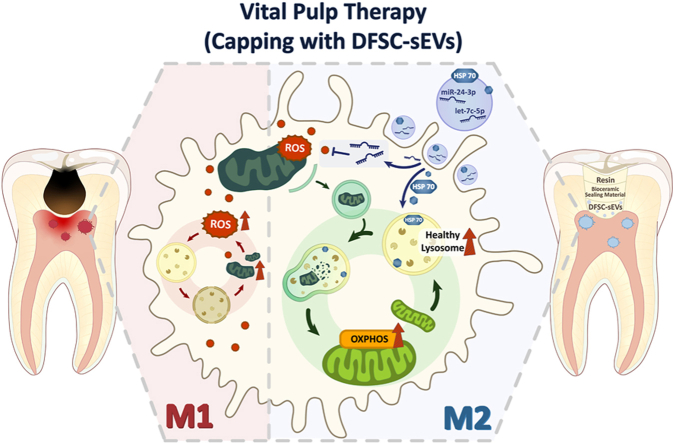
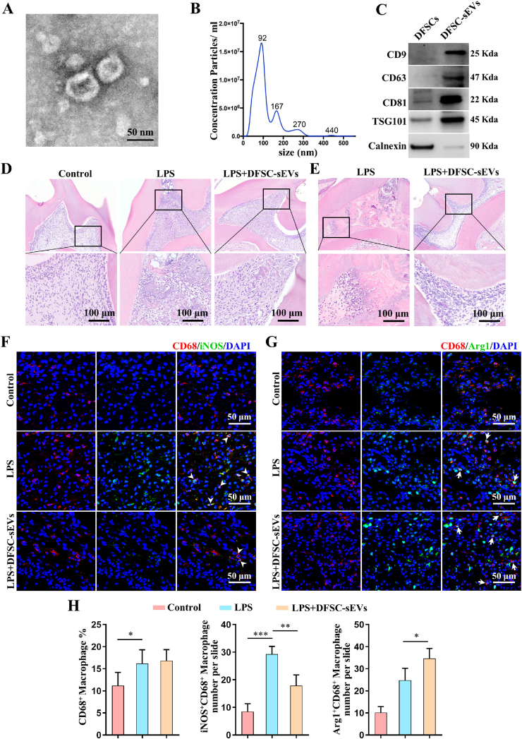
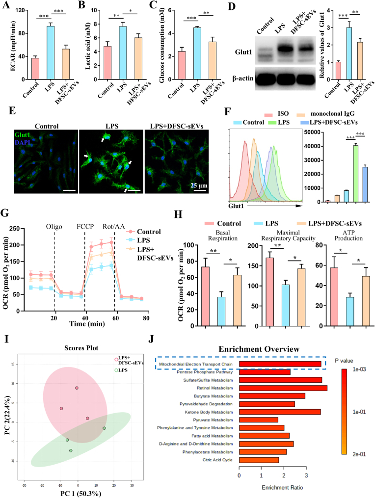
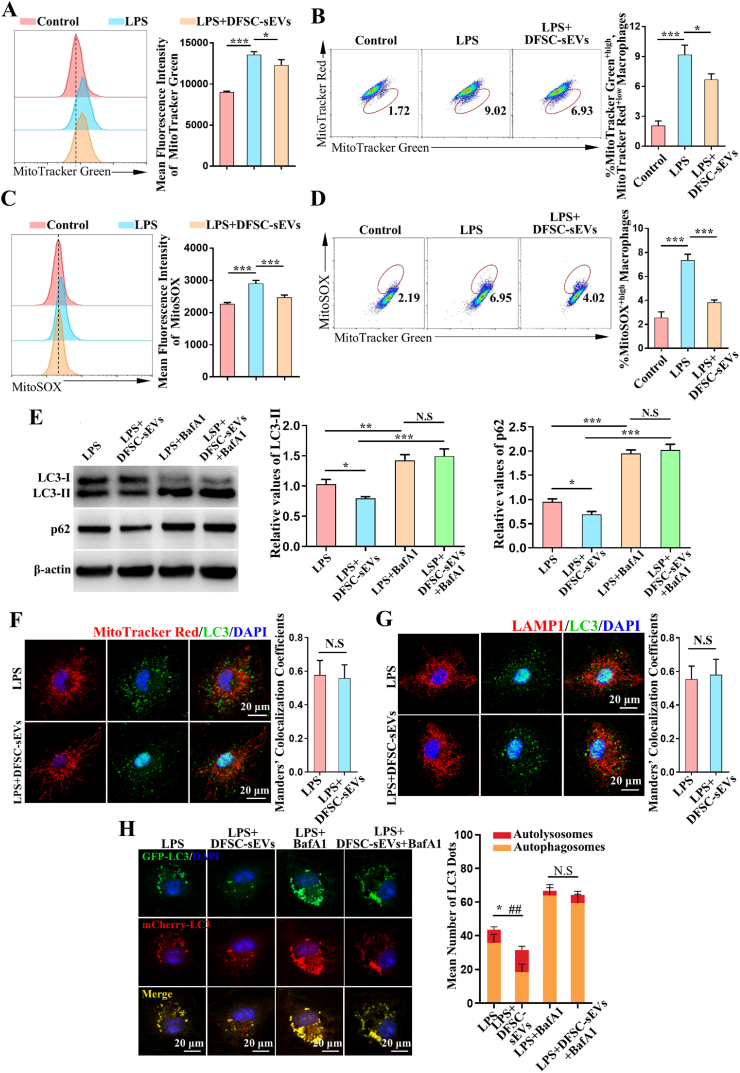
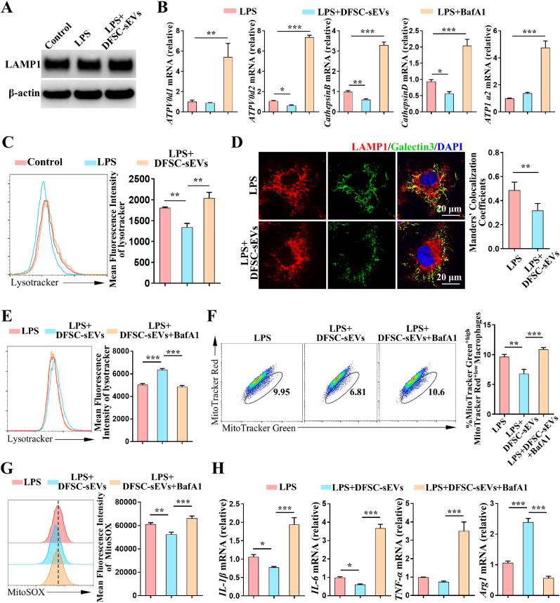
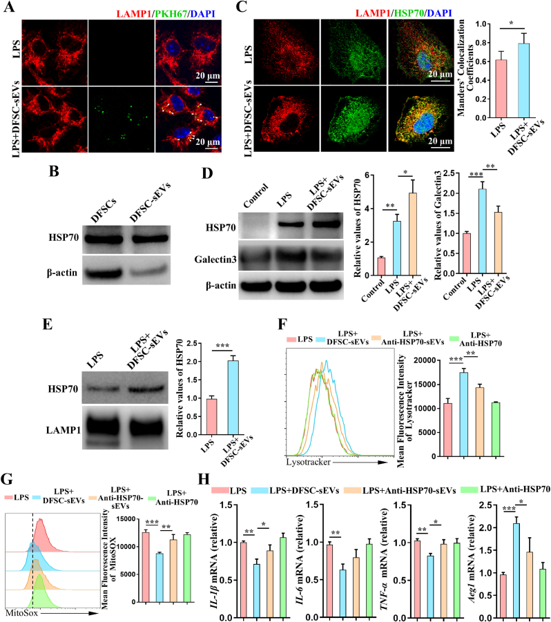
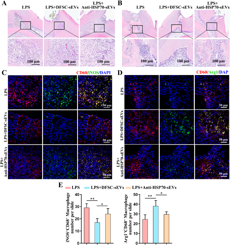
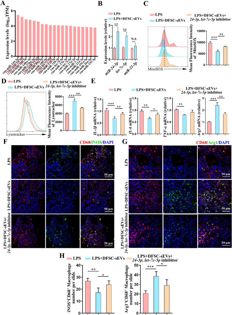
Vital pulp therapy (VPT) is considered a conservative means of preserving the vitality and function of the dental pulp after injury. However, current VPT has unfavorable effects on inflamed pulp. Mesenchymal stem cell (MSC)-derived small extracellular vesicles (MSC-sEVs) show powerful immunomodulatory capacities and exert therapeutic effects on a variety of inflammatory diseases. However, whether MSC-sEVs ameliorate the inflammatory response and promote inflammatory pulp repair in pulpitis is largely unknown. In this study, we show that sEVs derived from dental follicle stem cells (typical dental MSCs, DFSC-sEVs) alleviate lipopolysaccharide-induced pulpitis in rats and enhance pulp repair by inducing M2 macrophage polarization. Mechanistically, heat shock protein 70 (HSP70) within DFSC-sEVs can be supplemented into lysosomes to directly protect lysosomal function and induce mitophagy to promote the degradation of depolarized mitochondria, thereby preprogramming inflammatory macrophages to commit to oxidative phosphorylation, which fuels M2 polarization. Furthermore, DFSC-sEVs also transfer antioxidant miRNAs, including miR-24-3p and let-7c-5p, to inhibit mitochondrial reactive oxygen species production, thereby indirectly stabilizing lysosomes to induce M2 macrophage generation. Our study reveals a promising immunotherapeutic potential of DFSC-sEVs for VPT in inflamed pulp and a novel role for DFSC-sEVs in inhibiting the macrophage inflammatory response by protecting lysosomes and inducing mitophagy-mediated metabolic shifts toward oxidative phosphorylation.

Keywords: Dental follicle stem cell; Immunometabolism; Macrophage; Pulpitis; Small extracellular vesicle.


Conflict of interest statement

He Liu is an editorial board member for Bioactive Materials and was not involved in the editorial review or the decision to publish this article. Ya Shen is an associate editor for Bioactive Materials and was not involved in the editorial review or the decision to publish this article. All authors declare that there are no competing interests.

Figures

Image 1
Graphical abstract
Fig. 1
Fig. 1
DFSC-sEVs have therapeutic effects on LPS-induced pulpitis by inducing M2 macrophage polarization. (A) The ultrastructures of DFSC-sEVs were observed via transmission electron microscopy (TEM). (B) The size distribution of purified DFSC-sEVs was assessed via nanoparticle tracking analysis (NTA). (C) The expression of the sEV-associated protein markers CD9, CD63, CD81, TSG101 and Calnexin in DFSC-sEVs and DFSCs was assessed by western blotting. (D) H&E staining showing inflammatory cell infiltration in rats with LPS-induced pulpitis in rats at 3 d after capping with DFSC-sEVs (n = 5). (E) H&E staining showing pulpal repair in rats with LPS-induced pulpitis at 28 d after capping with DFSC-sEVs. CD68+iNOS+ M1 macrophages (white arrowhead) (F) and CD68+Arg1+ M2 macrophages (white arrow) (G) in the pulp after treatment with DFSC-sEVs were observed via laser-scanning confocal microscopy. (H) Statistical analysis of the ratio of CD68+ macrophages among all cells and the numbers of CD68+iNOS+ M1 macrophages and CD68+Arg1+ M2 macrophages. Error bars represent the mean ± s.d. ∗∗∗P < 0.005; ∗∗P < 0.01; and ∗P < 0.05.
Fig. 2
Fig. 2
DFSC-sEV-treated macrophages undergo metabolic reprogramming and commit to OXPHOS, which results in an anti-inflammatory phenotype. LPS-treated BMDMs were stimulated with or without DFSC-sEVs for 12 h. (A) The extracellular acidification rate (ECAR) of LPS-treated BMDMs under basal conditions was assessed. Lactate production (B) and glucose consumption (C) by BMDMs were determined using the culture supernatant. (D) The expression of Glut1 was determined by western blotting. (E) Glut1 expression and cell surface translocation (white arrow) were assessed via laser-scanning confocal microscopy. (F) The expression or fluorescence intensity of Glut1 on the surface of BMDMs was assessed via flow cytometry. (G) The OCRs of BMDMs at baseline and in response to sequential treatment with oligomycin (Oligo), FCCP, and rotenone plus antimycin A (Rot/AA) were measured. (H) Basal respiration, ATP production, and maximum respiratory capacity of the macrophages were analyzed. (I) Principal component analysis (PCA) plot of metabolomics data from LPS-treated BMDMs in the presence or absence of DFSC-sEVs (n = 4). (J) Metabolite set enrichment analysis (MSEA) of differentially abundant metabolites in LPS + DFSC-sEV-treated macrophages versus LPS-treated macrophages was performed using MetaboAnalyst. Error bars represent the mean ± s.d. ∗∗∗P < 0.005; ∗∗P < 0.01; and ∗P < 0.05.
Fig. 3
Fig. 3
DFSC-sEVs prevent the accumulation of dysfunctional mitochondria and the production of mitochondrial ROS in inflammatory macrophages by accelerating autophagic flux. LPS-treated BMDMs were stimulated with or without DFSC-sEVs for 12 h. Then, the BMDMs were labeled with mitochondrial membrane potential (ΔΨm)-independent MitoTracker Green and ΔΨm-sensitive MitoTracker Red probes. (A) Total mitochondrial mass in BMDMs was assessed by flow cytometry by analyzing the mean fluorescence intensity of MitoTracker Green. (B) The ratio of dysfunctional mitochondria (MitoTracker Green + high, MitoTracker Red + low) was assessed. (C) Treated BMDMs were labeled with MitoTracker Green and the mitochondria-specific ROS indicator MitoSOX. mtROS levels in BMDMs were determined by MitoSOX fluorescence intensity. (D) The ratio of mtROS production in BMDMs (MitoTracker Green + high, MitoSOX + high) was analyzed. (E) LPS-treated BMDMs were stimulated with or without DFSC-sEVs in the absence or presence of bafilomycin A1 (BafA1, 100 nM), which was added 2 h before the cells were harvested. The expression of LC3-I/II and p62 was determined by western blotting. The histogram shows the quantification of LC3-II and p62 expression after normalization to β-actin expression. (F) The colocalization of mitochondria (labeled with MitoTracker Red) with LC3 in BMDMs exposed to LPS and DFSC-sEVs was visualized via laser-scanning confocal microscopy. (G) The colocalization of LC3 puncta with lysosomes (labeled with LAMP1) was visualized using laser-scanning confocal microscopy in LPS-treated BMDMs in the presence or absence of DFSC-sEVs. (H) BMDMs were transfected with a lentiviral plasmid encoding mCherry-GFP-LC3B, and autophagic flux was assessed after treatment with LPS or DFSC-sEVs for 12 h. The graph on the right shows the quantification of LC3 puncta (∗P < 0.05 for total LC3 puncta; ##P < 0.01 for red puncta). The red puncta represent autolysosomes (with loss of GFP fluorescence in the acidic lysosomal environment). Error bars represent the mean ± s.d. ∗∗∗P < 0.005; ∗∗P < 0.01; and ∗P < 0.05.
Fig. 4
Fig. 4
DFSC-sEVs induce mitophagy by protecting lysosomal function. (A) LPS-treated BMDMs were stimulated with or without DFSC-sEVs for 12 h. The expression of LAMP1 was assessed by western blotting. (B) LPS-treated BMDMs were stimulated with or without DFSC-sEVs in the absence or presence of bafilomycin A1 (BafA1, 100 nM). The relative mRNA levels of the lysosome-associated genes ATP6V0d1, ATP6V0d2, CathepsinB, CathepsinD and ATP1A2 were determined via qPCR. (C) Lysosomal activity in BMDMs was assessed by flow cytometry by analyzing the mean fluorescence intensity of LysoTracker in BMDMs treated with LPS and DFSC-sEVs. (D) The colocalization of LAMP1 with galectin-3 was observed via laser-scanning confocal microscopy. LPS-treated BMDMs were stimulated with or without DFSC-sEVs in the absence or presence of BafA1. (E) The mean fluorescence intensity of LysoTracker in BMDMs was recorded. (F) The ratio of dysfunctional mitochondria was assessed in BMDMs labeled with MitoTracker Green and MitoTracker Red. (G) mtROS levels in BMDMs were determined using MitoSOX. (H) qPCR analysis of the relative mRNA levels of the macrophage polarization-related factors IL-1β, IL-6, TNF-α, and Arg1 in BMDMs. Error bars represent the mean ± s.d. ∗∗∗P < 0.005; ∗∗P < 0.01; and ∗P < 0.05.
Fig. 5
Fig. 5
DFSC-sEVs transfer HSP70 to stabilize lysosomes. (A) LPS-treated BMDMs were treated with PKH67-DFSC-sEVs for 12 h in vitro. The colocalization of DFSC-sEVs and LAMP1 (white arrow) was observed via laser-scanning confocal microscopy. (B) The expression of HSP70 in DFSCs and DFSC-sEVs was assessed by western blotting. LPS-treated BMDMs were stimulated with or without DFSC-sEVs for 12 h. (C) Immunofluorescence analysis showing the distribution of HSP70. (D) The expression of HSP70 and galectin-3 was determined by western blotting. (E) Western blot showing the levels of HSP70 in isolated lysosomes from BMDMs exposed to LPS and DFSC-sEVs. LPS-treated BMDMs were stimulated with DFSC-sEVs alone or with DFSC-sEVs and then incubated with a neutralizing antibody against HSP70 for 12 h. (F) The mean fluorescence intensity of LysoTracker in BMDMs was recorded. (G) mtROS levels were determined in BMDMs labeled with MitoSOX. (H) qPCR analysis of the relative mRNA levels of the macrophage polarization-related factors IL-1β, IL-6, TNF-α, and Arg1 in BMDMs. Error bars represent the mean ± s.d. ∗∗∗P < 0.005; ∗∗P < 0.01; and ∗P < 0.05.
Fig. 6
Fig. 6
HSP70 is required for the therapeutic effects of DFSC-sEVs on LPS-induced pulpitis. LPS-induced pulpitis in rats was capped with DFSC-sEVs alone or with neutralizing antibody against HSP70 (n = 5). (A) H&E staining showing inflammatory cell infiltration in LPS-induced pulpitis in rats 3 d after treatment. (B) H&E staining showing pulpal repair in LPS-induced pulpitis in rats at 28 d. CD68+iNOS+ M1 macrophages (C) and CD68+Arg1+ M2 macrophages (D) in the pulp were analyzed via laser-scanning confocal microscopy. (E) Statistical analysis of the numbers of CD68+iNOS+ M1 macrophages and CD68+Arg1+ M2 macrophages. Error bars represent the mean ± s.d. ∗∗∗P < 0.005; ∗∗P < 0.01; and ∗P < 0.05.
Fig. 7
Fig. 7
DFSC-sEVs transfer miR-24-3p and let-7c-5p to inhibit mtROS production, thereby protecting lysosomal function. (A) The histogram shows the top 20 miRNAs in DFSC-sEVs. (B) qPCR analysis showing the levels of miR-24-3p, let-7c-5p and miR-21-5p in LPS-treated BMDMs exposed to DFSC-sEVs. LPS-treated BMDMs were stimulated with or without DFSC-sEVs in the absence or presence of miR-24-3p and let-7c-5p inhibitors. (C) mtROS levels in BMDMs were determined using MitoSOX. (D) The mean fluorescence intensity of LysoTracker in BMDMs was recorded. (E) qPCR analysis of the relative mRNA levels of the macrophage polarization-related factors IL-1β, IL-6, TNF-α, and Arg1 in BMDMs. LPS-induced pulpitis in rats was induced by capping teeth with DFSC-sEVs and antagomirs of miR-24-3p and let-7c-5p (n = 5). CD68+iNOS+ M1 macrophages (F) and CD68+Arg1+ M2 macrophages (G) in the pulp were analyzed via laser-scanning confocal microscopy. (H) Statistical analysis of the numbers of CD68+iNOS+ M1 macrophages and CD68+Arg1+ M2 macrophages. ∗∗∗P < 0.005; ∗∗P < 0.01; and ∗P < 0.05.

Similar articles

References

    1. Duncan H.F., Galler K.M., Tomson P.L., Simon S., El-Karim I., Kundzina R., Krastl G., Dammaschke T., Fransson H., Markvart M., Zehnder M., Bjørndal L. European Society of Endodontology position statement: management of deep caries and the exposed pulp. Int. Endod. J. 2019;52:923–934. doi: 10.1111/iej.13080. - DOI - PubMed
    1. Arora S., Cooper P.R., Friedlander L.T., Rizwan S., Seo B., Rich A.M., Hussaini H.M. Potential application of immunotherapy for modulation of pulp inflammation: opportunities for vital pulp treatment. Int. Endod. J. 2021;54:1263–1274. doi: 10.1111/iej.13524. - DOI - PubMed
    1. Quigley R.M., Kearney M., Kennedy O.D., Duncan H.F. Tissue engineering approaches for dental pulp regeneration: the development of novel bioactive materials using pharmacological epigenetic inhibitors. Bioact. Mater. 2024;40:182–211. doi: 10.1016/j.bioactmat.2024.06.012. - DOI - PMC - PubMed
    1. Kim D.-H., Jang J.-H., Lee B.-N., Chang H.-S., Hwang I.-N., Oh W.-M., Kim S.-H., Min K.-S., Koh J.-T., Hwang Y.-C. Anti-inflammatory and mineralization effects of ProRoot MTA and endocem MTA in studies of human and rat dental pulps in vitro and in vivo. J. Endod. 2018;44:1534–1541. doi: 10.1016/j.joen.2018.07.012. - DOI - PubMed
    1. Colombo J.S., Moore A.N., Hartgerink J.D., D'Souza R.N. Scaffolds to control inflammation and facilitate dental pulp regeneration. J. Endod. 2014;40:S6–S12. doi: 10.1016/j.joen.2014.01.019. - DOI - PMC - PubMed

LinkOut - more resources