Comparative Study
. 2025 May 22:16:1547918.
doi: 10.3389/fimmu.2025.1547918. eCollection 2025.

Comparative analysis of neutrophil dynamics and disease in SARS-CoV-2 Delta and Omicron variants utilizing an in vivo feline model for COVID-19

Affiliations
Comparative Study

Comparative analysis of neutrophil dynamics and disease in SARS-CoV-2 Delta and Omicron variants utilizing an in vivo feline model for COVID-19

Sachithra Gunasekara et al. Front Immunol. .

Abstract

Introduction: The emergence of SARS-CoV-2 variants, particularly Delta (B.1.617.2) and Omicron (XBB.1.5) variants, has substantially influenced the clinical and immunological landscape of COVID-19. This study investigates the differential pathogenicity and immune responses in a feline model infected with these variants, focusing on neutrophil activation, neutrophil extracellular trap (NET) formation, and cytokine profiles.

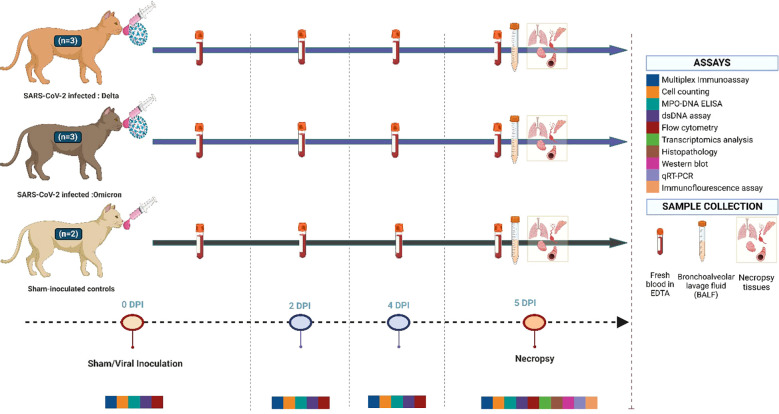
Methods: Eight pathogen-free cats were inoculated with B.1.617.2 (Delta) SARS-CoV-2 (n=3), XBB.1.5 (Omicron) SARS-CoV-2 (n=3), or vehicle (n=2), and clinical assessments, histopathological examinations, and cytokine analyses were performed post-infection.

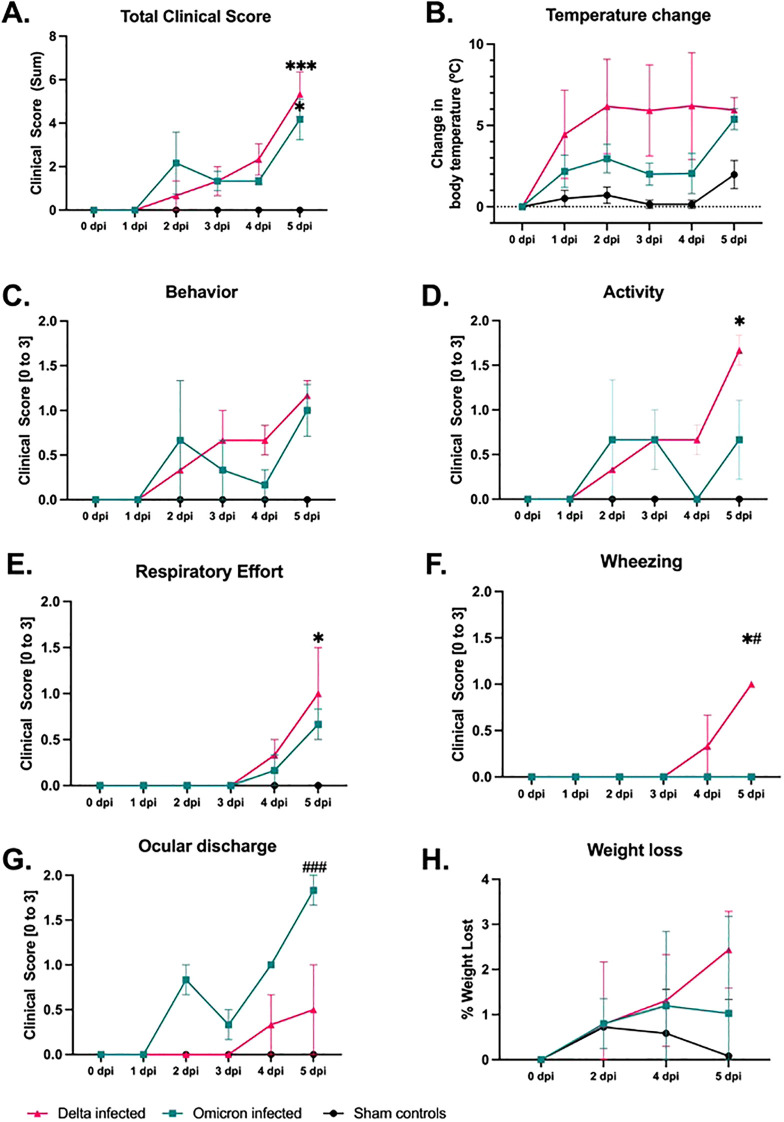
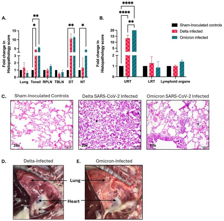
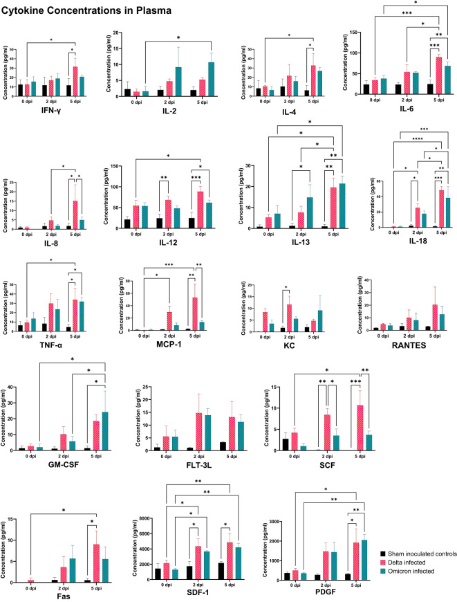
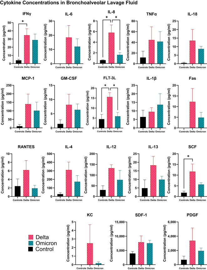
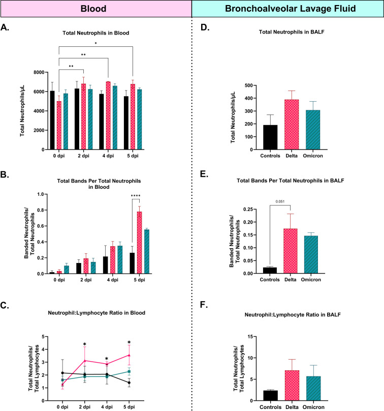
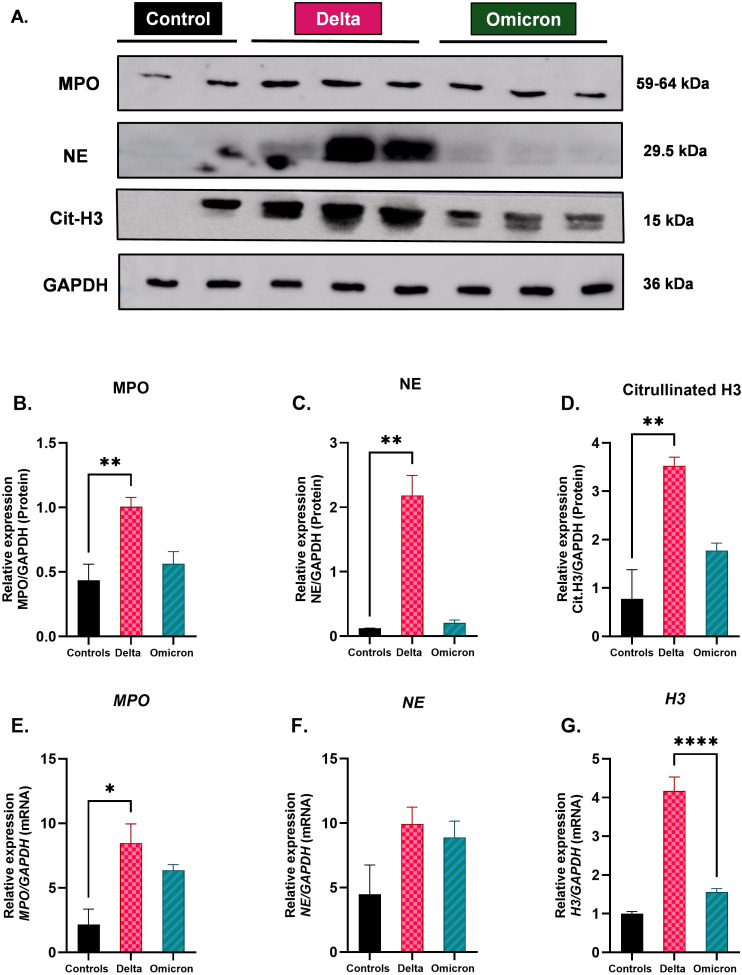
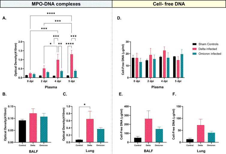
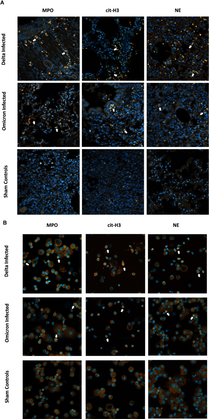
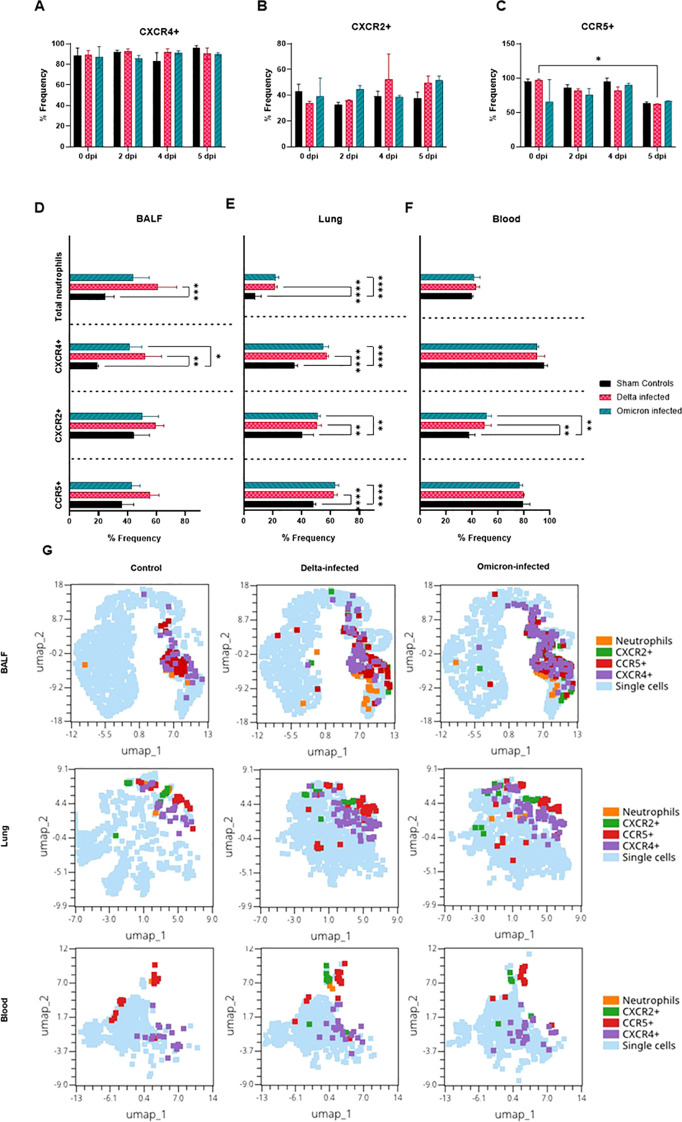
Results: Results demonstrate that Delta-infected cats exhibit more severe clinical manifestations characterized by significant elevation in respiratory effort, wheezing, and systemic inflammation compared to Omicron-infected cats, which show milder symptoms, primarily confined to the upper respiratory tract. Histopathological findings suggest pronounced lung damage in Delta-infected cats, whereas Omicron infection resulted in localized pathology. Cytokine profiling demonstrates heightened proinflammatory responses, particularly in Delta-infected cats, characterized by elevated levels of IL-6, IFN-γ and TNF-α while Omicron infection results in less pronounced inflammatory responses. Moreover, neutrophil-related parameters, including total neutrophil counts and banded neutrophils, were significantly elevated in Delta-infected cats, correlating with enhanced NET formation as evidenced by increased NETs-related markers MPO, NE, and citrullinated H3, and NET-specific markers MPO-DNA complexes and cell-free DNA.

Discussion: This study underscores the importance of variant-specific immune responses and highlights the need for targeted therapeutic strategies that mitigate severe lung injury associated with Delta infection, while also considering the distinct immune dynamics observed with the Omicron variant. Furthermore, results support the importance of delineating immune responses concerning future variants. These findings provide valuable insights into the pathogenesis of SARS-CoV-2 in companion animals and inform public health strategies as new variants continue to emerge.

Keywords: COVID-19; Delta; NETs; Omicron; SARS-CoV-2; feline; immunopathogenesis; neutrophils.


Conflict of interest statement

The authors declare that the research was conducted in the absence of any commercial or financial relationships that could be construed as a potential conflict of interest.

Figures

Figure 1
Figure 1
Experimental study design. This illustration outlines the experimental timeline, and the assays conducted throughout the study from a total of eight cats. Three were inoculated with the Delta (B.6.617.2) variant of SARS-CoV-2, three were inoculated with the Omicron (XBB.1.5) variant of SARS-CoV-2, with the remaining two serving as sham-inoculated controls. All cats were euthanized at 5 days post-infection (dpi) following necropsy. Whole blood, bronchoalveolar lavage, and tissues were collected for subsequent analysis. Image made in Biorender.
Figure 2
Figure 2
Comparative analyses of clinical parameters in cats infected with Delta or Omicron variants of SARS-CoV-2 and sham-inoculated controls. The figure illustrates the temporal progression of various clinical parameters recorded from a total of 8 cats: sham-inoculated controls (n=2), Delta-infected (n=3), and Omicron-infected (n=3). (A) Summated clinical scores for each group indicate significantly worsened clinical severity in all infected cats versus sham-inoculated controls. (B-H) provides the variation in individual clinical parameters. across the three groups over time: (B) Core temperature changes, (C) behavioral changes, (D) activity levels, (E) respiratory effort, (F) wheezing, (G) ocular discharge, and (H) percentage weight loss. Both variants of concern demonstrated significant disease progression compared to sham-inoculated controls, with Delta-infected cats causing the most severe overall progression. Statistical comparisons were conducted via two-way ANOVA and the data are represented as mean ± SEM. Statistical significances compared to sham controls are indicated by *p < 0.05, and ***p < 0.001 while comparisons between Delta versus Omicron variants are represented by #p < 0.05, and ### p < 0.001.
Figure 3
Figure 3
Pathological analysis of SARS-CoV-2 infected Delta and Omicron cats compared to sham controls. The figure illustrates both quantitative and qualitative analyses of tissue pathology following infection with different SARS-CoV-2 variants of concern. (A) represents quantitative histopathological scoring for each individual tissue (Lung, tonsil, retropharyngeal lymph node (RPLN), tracheobronchial lymph node (TBLN), distal trachea (DT), and nasal turbinate (NT). Both Delta and Omicron-infected groups exhibit significant changes in pathology scores across multiple tissues. (B) represents the quantitative histopathological scoring for grouped regions namely upper respiratory tract (URT), lower respiratory tract (LRT), and lymphoid organs. Both variants showed significantly higher scoring in the URT compared to the control group, with Omicron-infected cats exhibiting a notable increase compared to Delta-infected cats. (C) displays H&E stained lung sections from cats infected with Delta and Omicron variants of SARS-CoV-2, as well as sham-inoculated controls. Sham-inoculated controls exhibit intact alveolar architecture, with thin septa and clear alveolar spaces. Delta-infected lungs display the most severe architectural damage, including substantial septal thickening, heavy inflammatory infiltration of neutrophils and macrophages (black arrowhead), alveolar collapse, and consolidation, indicating extensive tissue damage and impaired lung function. In contrast, Omicron-infected lungs show mild septal thickening and moderate neutrophil infiltration (black arrow), with partial disruption of the alveolar structure. (D, E) demonstrates the gross pathology of the lungs from SARS-CoV-2 (D) Delta and (E) Omicron-infected cats. Images highlight the greater severity of lung pathology in Delta-infected cats compared to those infected with the Omicron variant. Statistical significance was assessed using two-way ANOVA, and p-values are indicated as follows: *p < 0.05, **p < 0.01, ****p < 0.0001 with data represented as mean ± SEM. Magnification: (C) 20x, scale bar = 100 µm.
Figure 4
Figure 4
Cytokine and chemokine profiles in plasma of cats infected with SARS-CoV-2 Delta and Omicron variants. This figure demonstrates cytokine concentrations in plasma collected at 0 dpi, 2 dpi, and 5 dpi from all eight cats. Significant alterations were observed in both variants throughout the time points compared to sham inoculated controls. Delta-infected cats showed higher expression levels of several key proinflammatory mediators including IFN-γ, IL-6, IL-8, and TNF-α, particularly at 5 dpi. Statistical analyses were performed using one-way ANOVA and data are presented as mean ± SEM (*p < 0.05, **p < 0.01, ***p < 0.001, ****p < 0.0001).
Figure 5
Figure 5
Cytokine and chemokine profiles in BALF of cats infected with SARS-CoV-2 Delta and Omicron variants. This figure illustrates the cytokine concentrations in BALF collected at 5 dpi from all cats. Significant alterations were observed in both variants compared to sham inoculated controls with the Delta-infected cats demonstrating a more pronounced increase in all the cytokines.Statistical analyses were performed using one-way ANOVA and data are presented as mean ± SEM (*p < 0.05).
Figure 6
Figure 6
Enrichment in Gene Ontology (GO) terms and Kyoto Encyclopedia of Genes and Genomes (KEGG) pathway analyses of differentially expressed genes (DEGs) in SARS-CoV-2 infected cats compared to controls, and between Delta and Omicron variants. GO and KEGG pathway enrichment analyses show the differential molecular response of cats infected with Delta and Omicron variants of SARS-CoV-2. (A) Bar plots of enriched GO terms, (B) Bubble plot of top enriched KEGG pathways for DEGs from Delta-infected cats compared to sham-inoculated controls. (C) Bar plots of enriched GO terms, (D) Bubble plot of top enriched KEGG pathways for DEGs from Omicron-infected cats compared to sham-inoculated controls. (E) Bar plots of enriched GO terms, (F) Bubble plot of top enriched KEGG pathways for DEGs from Delta-infected cats compared to Omicron-infected cats.
Figure 7
Figure 7
Comparative assessment of neutrophil-related parameters, including total neutrophil counts, neutrophil-to-lymphocyte ratio (NLR), and banded neutrophil counts in cats infected with Delta and Omicron variants of SARS-CoV-2, with sham controls. (A-C) demonstrates neutrophil counts, banded neutrophils, and NLR obtained from blood smears prepared from all eight cats throughout the study at 0 dpi, 2 dpi, 4 dpi, and 5 dpi while (D-F) provides neutrophil counts, banded neutrophils, and NLR obtained from BALF of all cats at 5 dpi, respectively. Delta-infected cats indicated an increase in all three parameters in both blood and BALF with significant alterations in total neutrophil counts of blood. Statistical comparisons were performed using two-way and one-way ANOVA and data is represented as mean ± SEM (*p < 0.05, **p < 0.01, ****p < 0.0001).
Figure 8
Figure 8
Evaluation of expression in neutrophil activation and NETs-related markers in cats infected with Delta and Omicron variants of SARS-CoV-2. (A) Representative western blot image of MPO, NE, and citrullinated histone H3 in lung tissues of all eight cats. GAPDH serves as a loading control. (B-D) Relative protein expression levels for MPO, NE, and Cit-H3 normalized to GAPDH, respectively. All three markers were significantly increased in Delta-infected cats. (E-G) Quantitative RT-PCR demonstrating mRNA expression levels of MPO, NE, and histone H3 (H3), normalized to GAPDH, respectively. mRNA levels were significantly upregulated in MPO of Delta-infected cats compared to controls and H3 compared to Omicron-infected cats. Statistical interpretations were performed using one-way ANOVA. Data are presented as mean ± SEM; statistical significance is indicated by asterisks: *p < 0.05, **p < 0.01, ****p < 0.0001.
Figure 9
Figure 9
Concentrations of MPO-DNA complexes and cell-free DNA in blood, BALF, and lung tissues obtained from all cats over time. (A) MPO-DNA complexes in plasma (B) MPO-DNA complexes in BALF and (C) MPO-DNA complexes in lung tissue revealed significant alterations in MPO-DNA complexes favoring Delta-infected cats at 2, 4, and 5 dpi, as compared to the controls and Omicron-infected cats. Similarly, (D) cell-free DNA in blood, (E) cell-free DNA in BALF, and (F) cell-free DNA in lungs were altered majorly in Delta-infected cats compared to Omicron-infected cats and sham-inoculated controls. The statistical analyses were performed using two-way and one-way ANOVA. Data are represented as mean ± SEM (*p < 0.05, **p < 0.01, ***p < 0.001, ****p < 0.0001).
Figure 10
Figure 10
Visual representation of NETs-related markers in lung tissue and BALF in cats following SARS-CoV-2 Infection. Immunofluorescence staining was performed on lung tissues and BAL cells from all cats localizing myeloperoxidase (MPO), citrullinated H3 (cit-H3), and neutrophil elastase (NE). (A) Immunofluorescence images from lung tissues of cats infected with SARS-CoV-2 variants of concern (Delta or Omicron) compared to sham-inoculated controls. (B) Immunofluorescence images from cytospin slides of BAL from the same groups. DAPI was used as the nuclei counter stain highlighting DNA in blue while MPO/NE/citrullinated H3 was stained in orange. White arrows indicate active neutrophils potentially involved in NETs formation. Magnification (A, B): 20x, scale bar = 50 µm.
Figure 11
Figure 11
Flow cytometric analysis of neutrophil subsets across blood, BALF, and lung tissues in SARS-CoV-2 infected cats (Delta, Omicron) and controls. (A-C) Bar charts represent neutrophil subsets in blood expressing (A) CXCR4+, (B) CXCR2+, and (C) CCR5+ at different time points (0, 2,4, 5 dpi for Delta and Omicron-infected cats compared to controls. (D-F) CXCR4+, CXCR2+, CCR5+, and total neutrophils in BALF, lung tissues, and blood at 5 dpi, respectively. (G) UMAP plots showing the clustering of neutrophil subsets (CXCR4+, CXCR2+, CCR5+) in BALF, lung, and blood, with different subsets highlighted in respective colors (neutrophils, CXCR2+, CCR5+, CXCR4+). Statistical significance is indicated for key findings using two-way and one-way ANOVA and data are represented as mean ± SE (*p < 0.05, **p < 0.01, ***p < 0.001, ****p < 0.0001).

Similar articles

References

    1. Aleem A, Ab AS, Slenker AK. Emerging variants of SARS-CoV-2 and novel therapeutics against coronavirus (COVID-19). (2021). - PubMed
    1. Fontanet A, Autran B, Lina B, Kieny MP, Karim SSA, Sridhar D. SARS-CoV-2 variants and ending the COVID-19 pandemic. Lancet. (2021) 397:952–4. doi: 10.1016/S0140-6736(21)00370-6 - DOI - PMC - PubMed
    1. Maurya R, Swaminathan A, Shamim U, Arora S, Mishra P, Raina A, et al. . Co-evolution of SARS-CoV-2 variants and host immune response trajectories underlie COVID-19 pandemic to epidemic transition. Iscience. (2023) 26. doi: 10.1016/j.isci.2023.108336 - DOI - PMC - PubMed
    1. Tulimilli SV, Dallavalasa S, Basavaraju CG, Kumar Rao V, Chikkahonnaiah P, Madhunapantula SV, et al. . Variants of severe acute respiratory syndrome coronavirus 2 (SARS-CoV-2) and vaccine effectiveness. Vaccines. (2022) 10:1751. doi: 10.3390/vaccines10101751 - DOI - PMC - PubMed
    1. Zabidi NZ, Liew HL, Farouk IA, Puniyamurti A, Yip AJW, Wijesinghe VN, et al. . Evolution of SARS-CoV-2 variants: implications on immune escape, vaccination, therapeutic and diagnostic strategies. Viruses. (2023) 15:944. doi: 10.3390/v15040944 - DOI - PMC - PubMed

Publication types

Supplementary concepts