Review
. 2025 May 22:13:1569337.
doi: 10.3389/fcell.2025.1569337. eCollection 2025.

Organoids technology in cancer research: from basic applications to advanced ex vivo models

Affiliations
Review

Organoids technology in cancer research: from basic applications to advanced ex vivo models

Luca Varinelli et al. Front Cell Dev Biol. .

Abstract

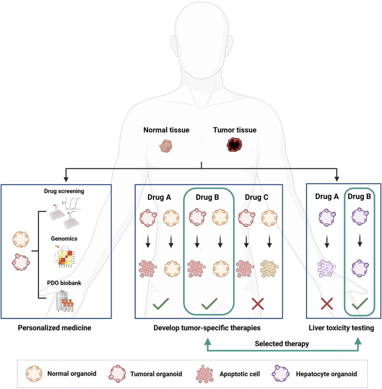
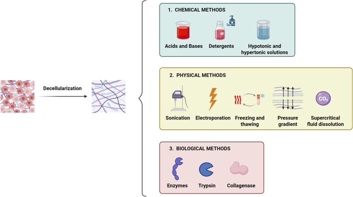
Patient-derived organoids (PDOs) are tridimensional cultures derived from the stem component of a tissue. They preserve the genetic and phenotypic characteristics of the tissue of origin, and represent valuable in vitro models for drug screening, biomarker discovery, cell therapy and genetic modification. Importantly, PDOs reproduce the tumor behavior and can predict therapeutic responses, making them relevant for clinical applications for personalized therapies. PDOs may also be used for studying the interactions between cancer cells and the tumor microenvironment (TME). These interactions are driven by biochemical factors released by the cells, and biomechanical events such as the remodeling of the extracellular matrix (ECM). In recent years, it has become evident that the interactions between cancer cells and the TME have an impact on tumor development and on the efficacy of cancer therapy Therefore, targeting both tumor cells and the TME may improve patient response to treatment. Most PDO culture protocols are limited to epithelial cells. However, recent advances such as use of decellularized ECM (dECM) scaffolds have allowed for the development of in vivo-like environments that host diverse cell types, both normal and pathological, in a tridimensional (3D) manner that closely mimics the complexity of the TME. dECM-based models effectively replicate the interactions between tumor cells, ECM and the microenvironment, are easy to analyze and adaptable for drug testing. By incorporating TME components and therapeutic agents, these models offer an advanced platform for preclinical testing.

Keywords: cancer organoids; decellularized matrix; drug screening; ex vivo cancer models; extracellular matrix (ECM); personalized therapy.


Conflict of interest statement

The authors declare that the research was conducted in the absence of any commercial or financial relationships that could be construed as a potential conflict of interest.

Figures

FIGURE 1
FIGURE 1
This figure summarizes the main methods used for organoid culture. Organoids can be developed from tissue-derived cells (TDCs), from adult induced-pluripotent stem cells (iPSCs) or from organ-restricted adult stem cells (aSCs). Depending on the source of the cells used to generate the organoids, the growth and expansion media need to be supplemented with growth factors and/or specific soluble factors. Organoids are grown in matrices of either animal or synthetic origin, which provide structural support and promote cell aggregation in a 3D manner. Organoids can be developed, grown and expanded using advanced culture techniques, such as Organ-on-a-chip technology, 3D bioprinting, and various co-culture methods (i.e., Air-liquid-interface system, cell-culture insert). Key features of the different organoid culture methods are highlighted in the figure (see boxes). Legend: TDCs, Tissue-derived Cells; iPSCs, induced-Pluripotent Stem Cells; BMP4, Bone Morphogenetic Protein 4; Wnt, Wnt protein; FGF, Fibroblast Growth Factor; VEGF, Vascular Endothelial Growth Factor; BMP, Bone Morphogenic Protein; TGF, Transforming Growth Factor; ECM, Extracellular Matrix.
FIGURE 2
FIGURE 2
Schematic representation of the different organoids that can be developed from iPSC and aSC cells, along with the various growth factors required for their development. The signaling components essential for guided differentiation and niche function are shown, with activated signaling pathways shown in green, and inhibited ones in red. Key factors include BMP, bone morphogenetic protein; EGF, epidermal growth factor; FGF, fibroblast growth factors; HGF, hepatocyte growth factor; IGF, insulin-like growth factor; ROCK, RHO-associated protein kinase; TGF, transforming growth factor; VEGF, vascular endothelial growth factor.
FIGURE 3
FIGURE 3
3D PDO cultures derived from various biobanks, which mimic the key features of different cancer types, are used for drug screening to identify potential therapeutic markers that could help personalize patient treatment.
FIGURE 4
FIGURE 4
How organoids can be used for personalized cancer treatment and drug development. Organoids are developed from patient-derived tissue, both healthy and cancerous. Once developed, organoids can be characterized from a genetic point of view and used in drug screenings to correlate the genetic landscape of the tumor with the pharmacological response. The development of healthy organoids makes it possible to select less toxic drugs by searching for compounds that can selectively kill cancer cells only. Furthermore, organoids derived from healthy liver tissue can be used to test the hepatotoxicity of new drugs.
FIGURE 5
FIGURE 5
Modulation of the main hallmarks of cancer by the ECM. The remodeling of the ECM during cancer development allows the formation of bonds between the proteins and the molecules that make up the matrix, such as collagen, fibronectin and laminin with different receptors on the cell surface. These interactions activate intracellular signaling pathways that promote key pro-tumor actions, including survival, proliferation, angiogenesis, metastasis and resistance to chemotherapy. Both neoplastic cells and the non-malignant stromal cells contribute to those processes and, in turn, are influenced by changes in the matrix. These modifications encompass i) biochemical changes, ii) secretion of specific growth factors, iii) alterations in matrix hydration, iv) post-translational modifications, v) changes in biomechanical properties, vi) massive collagen deposition leading to a more fibrotic state, vii) alterations in the structural organization with changes in ECM porosity, viii) deregulated turnover rates of matrix components and ix) disruption of cell-cell adhesion interactions due to expression of binding proteins.
FIGURE 6
FIGURE 6
Schematic illustration of the role and functions of extracellular matrix in the tumor microenvironment. Summary of the main effects and key cell-matrix interactions involving ECM molecules in various types of cancer. 1) Biochemical and biophysical interactions, mediated by cell-ECM communication through specific receptors on the ECM (e.g., integrins), promote the expression of collagen, tenascin-c and, in some cases, proteoglycans, favoring tumor growth. 2) ECM remodeling results in an accumulation of structural proteins (such as collagen and proteoglycans), at the tumor site, forming a protective barrier against drugs and inhibiting immune cells activity. Additionally, ECM remodeling activates signaling pathways that regulate cell-ECM interaction via specific receptors (i.e., integrins and Toll-like receptors). 3) The overexpression of structural and ECM-related proteins enhances tumor invasion. 4) The overexpression of periostin and tenascin-c, driven by remodeling activity of neoplastic cells and the TME, contributes to the formation of the metastatic niche. Legend, ECM, Extracellular matrix; CAF, Cancer-Associated Fibroblast; TLRs, Toll-like Receptors; TME, Tumor Microenvironment.
FIGURE 7
FIGURE 7
Main methods used for tissue decellularization. The decellularization process involves the complete removal of the cellular component from the tissue while preserving the native ECM micro-architecture and biochemical properties. The main decellularization methods are categorized into: i) chemical type, ii) physical type and iii) biological type. Often, combining different methods enhances the efficiency of decellularization.
FIGURE 8
FIGURE 8
The figure illustrates the general process for obtaining decellularized tissues to be used as scaffolds for organoid culture and/or different cellular subpopulations. The tissue undergoes several washing cycles with non-ionic detergents, until the complete removal of cellular material from the native tissue. The decellularization process can take from hours to days, depending on the tissue’s size and origin. Once decellularized, the tissue can be used in its lyophilized and naïve form for the culture of organoids and/or other cell types within the microenvironment. 1) lyophilized dECM can be used to produce biocompatible inks for 3D bioprinting of scaffolds, which can then be repopulated with organoids. 2) Lyophilized dECM can be resuspended at various concentrations in synthetic hydrogels and used as support matrices for organoid growth as an alternative to Matrigel. 3) Lyophilized dECM can be resuspended, at different concentrations, directly inside the organoid culture medium. 4) Naïve dECM can be used directly as scaffolds and repopulated with organoids. Legend: dECM, decellularized Extracellular Matrix; PBS, Phosphate Buffer Saline; SDS, Sodium Dodecyl Sulfate.
FIGURE 9
FIGURE 9
Overview of the main sources from which dECMs are obtained and of the main methodologies used to obtain them, including the various modelling strategies to develop 3D disease models that can summarize the main features of a tumor.
FIGURE 10
FIGURE 10
Overview of the main strategies used to develop 3D in vitro tumor models based on dECM. The main advantages and limitations of all strategies are also indicated.

Similar articles

Cited by

References

    1. Aggarwal D., Russo S., Naik P., Bhatia S., Spector D. L. (2023). Establishment and culture of patient-derived breast organoids. J. Vis. Exp. 17 (192). 10.3791/64889 - DOI - PMC - PubMed
    1. Akhtar M., Haider A., Rashid S., Dakhilalla A., Al-Nabet A. D. M. H. (2019). Paget’s “seed and soil” theory of cancer metastasis: an idea whose time has come. Adv. Anat. Pathol. 26, 69–74. 10.1097/PAP.0000000000000219 - DOI - PubMed
    1. Alcaraz J., Otero J., Jorba I., Navajas D. (2018). Bidirectional mechanobiology between cells and their local extracellular matrix probed by atomic force microscopy. Semin. Cell Dev. Biol. 73, 71–81. 10.1016/j.semcdb.2017.07.020 - DOI - PubMed
    1. Alessandrini A., Facci P. (2005). AFM: a versatile tool in biophysics. Meas. Sci. Technol. 16, R65–R92. 10.1088/0957-0233/16/6/R01 - DOI
    1. Alsaed B., Smolander J., Laitinen H., Lin L., Bobik N., Lahtinen L., et al. (2024). Ex vivo modeling of precision immuno-oncology responses in lung cancer. Sci. Adv. 10, eadq6830. 10.1126/sciadv.adq6830 - DOI - PMC - PubMed

LinkOut - more resources