. 2025 May 22:13:1494660.
doi: 10.3389/fcell.2025.1494660. eCollection 2025.

Dietary modifications affect renal recovery during the healing phase following ischemic acute ischemic kidney injury

Affiliations

Dietary modifications affect renal recovery during the healing phase following ischemic acute ischemic kidney injury

Junseok Jeon et al. Front Cell Dev Biol. .

Abstract

Introduction: The effects of dietary modifications, such as varying amounts of salt, fat, and protein intake on the healing phase of acute kidney injury (AKI) remain to be elucidated. We investigated the effects of low- or high-salt/fat/protein diets on the intrarenal immunologic micromilieu and healing after renal ischemia-reperfusion injury (IRI) using murine ischemic AKI and human kidney-2 (HK-2) cell hypoxia models.

Methods: Three cohorts of male C57BL/6 mice (9-weeks old) were fed the designated diet from the third day following renal IRI until sacrifice (6 or 12 weeks after bilateral or unilateral IRI, respectively) in groups as follows: cohort 1, control, high- and low-salt/fat/protein; cohort 2, control, high- and low-salt; cohort 3, control, high- and low-fat/protein. Hypoxic HK-2 cells were treated with sodium chloride, amino acids, or fatty acids.

Results: Low-salt/fat/protein diet aggravated interstitial fibrosis, enhanced TGF-β expression, and induced more proinflammatory changes after bilateral IRI. High-salt diet aggravated renal tubular damage and enhanced the expression of intrarenal TGF-β after bilateral IRI, whereas low-salt diet enhanced the expression of intrarenal TGF-β after unilateral IRI. Low-salt diet induced more proinflammatory changes after bilateral IRI. Blood urea nitrogen levels were lower in the low fat/protein group than that in the control group following IRI. However, low-fat/protein diet aggravated interstitial fibrosis and enhanced intrarenal TGF-β expression after unilateral IRI. High sodium- or protein-containing media suppressed the proliferation of hypoxic HK-2 cells, whereas high lipid-containing media enhanced the proliferation of hypoxic HK-2 cells.

Conclusion: Excessive low or high salt, low fat, and low protein diet may adversely affect the healing process following renal IRI, supporting the importance of adequate and balanced nutrition during the recovery phase of ischemic AKI.

Keywords: acute kidney injury; diet; healing; immunologic micromilieu; ischemia-reperfusion injury.


Conflict of interest statement

The authors declare that the research was conducted in the absence of any commercial or financial relationships that could be construed as a potential conflict of interest.

Figures

FIGURE 1
FIGURE 1
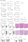
Changes in renal function, tubular injury, and fibrosis in mice fed high- or low-salt/fat/protein diet after renal ischemia-reperfusion injury. (A) Changes in body weight, blood pressure, renal function, and total cholesterol. (B) Tubular damage or atrophy. (C) Degree of interstitial fibrosis. (D) Intrarenal expression of TGF-β. *P < 0.05. Statistical analyses were performed using analysis of variance test followed by Tukey’s test.
FIGURE 2
FIGURE 2
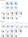
Leukocyte trafficking into the post-ischemic kidneys in mice fed low- or high-salt/protein/fat diet. Proportion of intrarenal total leukocytes expressing CD45 among total nuclei in the whole field after (A) bilateral and (B) unilateral ischemia-reperfusion injury. *P < 0.05. Statistical analyses were performed using analysis of variance test followed by Tukey’s test. Activated T cell, CD69+ subset; effector memory T cells, CD44+ CD62L-subset.
FIGURE 3
FIGURE 3
Intrarenal leukocyte subtypes in the post-ischemic kidneys in mice fed low- or high-salt/protein/fat diet. Intrarenal leukocyte subtypes after (A) bilateral and (B) unilateral ischemia-reperfusion injury. *P < 0.05. Statistical analyses were performed using analysis of variance test followed by Tukey’s test. Activated T cells, CD69+ subset; Effector memory T cells, CD44+ CD62L-subset; Regulatory CD4 T cells, FoxP3+ CD25+ CD4 T cell subset.
FIGURE 4
FIGURE 4
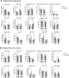
Expression of intrarenal cytokines and chemokines in the post-ischemic kidneys in mice fed low- or high-salt/protein/fat diet. Expression of intrarenal cytokines and chemokines after (A) bilateral and (B) unilateral ischemia-reperfusion injury. *P < 0.05. Statistical analyses were performed using analysis of variance test followed by Tukey’s test.
FIGURE 5
FIGURE 5
Changes in renal function, tubular injury, and fibrosis in mice fed low or high-salt diet after renal ischemia-reperfusion injury. (A) Changes in body weight, blood pressure, renal function, and total cholesterol. (B) Tubular necrosis, damage, or atrophy. (C) Degree of interstitial fibrosis. (D) Intrarenal expression of TGF-β. *P < 0.05. Statistical analyses were performed using analysis of variance test followed by Tukey’s test.
FIGURE 6
FIGURE 6
Leukocyte trafficking into the post-ischemic kidneys in mice fed low- or high-salt diet. Proportion of intrarenal total leukocytes expressing CD45 among total nuclei in the whole field after (A) bilateral and (B) unilateral ischemia-reperfusion injury. *P < 0.05. Statistical analyses were performed using analysis of variance test followed by Tukey’s test.
FIGURE 7
FIGURE 7
Intrarenal leukocyte subtypes in the post-ischemic kidneys in mice fed low- or high-salt diet. Intrarenal leukocyte subtypes after (A) bilateral and (B) unilateral ischemia-reperfusion injury. *P < 0.05. Statistical analyses were performed using analysis of variance test followed by Tukey’s test. Activated T cell, CD69+ subset; effector memory T cells, CD44+ CD62L-subset.
FIGURE 8
FIGURE 8
Expression of intrarenal cytokines and chemokines in the post-ischemic kidneys in mice fed low- or high-salt diet. Expression of intrarenal cytokines and chemokines after (A) bilateral and (B) unilateral ischemia-reperfusion injury. *P < 0.05. Statistical analyses were performed using analysis of variance test followed by Tukey’s test.
FIGURE 9
FIGURE 9
Changes in renal function, tubular injury, and fibrosis in mice fed high- or low-fat/protein diets after renal ischemia-reperfusion injury. (A) Changes in body weight, blood pressure, renal function, and total cholesterol. (B) Tubular necrosis, damage, or atrophy. (C) Degree of interstitial fibrosis. (D) Intrarenal expression of TGF-β. *P < 0.05. Statistical analyses were performed using analysis of variance test followed by Tukey’s test.
FIGURE 10
FIGURE 10
Leukocyte trafficking into the post-ischemic kidneys in mice fed low- or high-fat/protein diets. Proportion of intrarenal total leukocytes expressing CD45 among total nuclei in the whole field after (A) bilateral and (B) unilateral ischemia-reperfusion injury. *P < 0.05. Statistical analyses were performed using analysis of variance test followed by Tukey’s test.
FIGURE 11
FIGURE 11
Intrarenal leukocyte subtypes in the post-ischemic kidneys in mice fed low- or high-fat/protein diets. Intrarenal leukocyte subtypes after (A) bilateral and (B) unilateral ischemia-reperfusion injury. *P < 0.05. Statistical analyses were performed using analysis of variance test followed by Tukey’s test. Activated T cells, CD69+ subset; Effector memory T cells, CD44+ CD62L-subset; Regulatory CD4 T cells, FoxP3+ CD25+ CD4 T cell subset; Activated B cells, CD69+ B cell subset; Plasma cells, CD138+ CD126+ subset; Mature B cells, CD21/35 + B cell subset; Memory B cells, CD27+ B cell subset.
FIGURE 12
FIGURE 12
Expression of intrarenal cytokines and chemokines in the post-ischemic kidneys in mice fed low- or high-fat/protein diets. Expression of intrarenal cytokines and chemokines after (A) bilateral and (B) unilateral ischemia-reperfusion injury. *P < 0.05. Statistical analyses were performed using analysis of variance test followed by Tukey’s test.
FIGURE 13
FIGURE 13
Proliferation of hypoxic HK-2 cells treated with various salt, lipid, and protein concentrations. Proliferation of hypoxic HK-2 cells according to various (A) sodium concentrations and (B) protein or lipid concentrations in media. *P < 0.05. Statistical analyses were performed using analysis of variance test followed by Tukey’s test.

Similar articles

References

    1. Andrews P. M. (1990). Dietary protein regimen prior to renal ischemia significantly affects the postischemic uremic response. Nephron 55 (3), 272–276. 10.1159/000185974 - DOI - PubMed
    1. Andrews P. M., Bates S. B. (1986). Dietary protein prior to renal ischemia dramatically affects postischemic kidney function. Kidney Int. 30 (3), 299–303. 10.1038/ki.1986.184 - DOI - PubMed
    1. Ascon D. B., Lopez-Briones S., Liu M., Ascon M., Savransky V., Colvin R. B., et al. (2006). Phenotypic and functional characterization of kidney-infiltrating lymphocytes in renal ischemia reperfusion injury. J. Immunol. 177 (5), 3380–3387. 10.4049/jimmunol.177.5.3380 - DOI - PubMed
    1. Cao W., Li A., Wang L., Zhou Z., Su Z., Bin W., et al. (2015). A salt-induced reno-cerebral reflex activates renin-angiotensin systems and promotes CKD progression. J. Am. Soc. Nephrol. 26 (7), 1619–1633. 10.1681/asn.2014050518 - DOI - PMC - PubMed
    1. Chawla L. S., Eggers P. W., Star R. A., Kimmel P. L. (2014). Acute kidney injury and chronic kidney disease as interconnected syndromes. N. Engl. J. Med. 371 (1), 58–66. 10.1056/NEJMra1214243 - DOI - PMC - PubMed

LinkOut - more resources