

Sebaceous lymphadenoma of the parotid: a rare case report of an entity mimicking other salivary tumors
- PMID: 40476025
- PMCID: PMC12140100
- DOI: 10.1093/jscr/rjaf382
Sebaceous lymphadenoma of the parotid: a rare case report of an entity mimicking other salivary tumors
Abstract
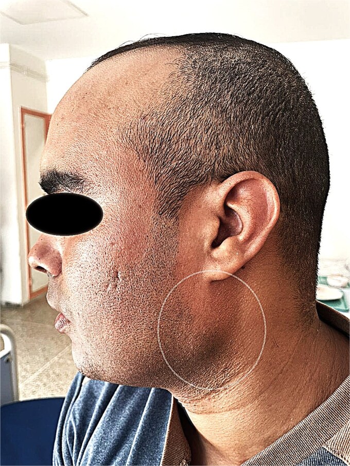
Sebaceous lymphadenoma is a rare benign tumor of the salivary glands, with fewer than 50 cases reported worldwide. Its clinical and radiological resemblance to malignant tumors poses diagnostic challenges. A 35-year-old female presented with a slow-growing, painless left parotid mass persisting for 2 years. Ultrasonography revealed a well-circumscribed, hypoechoic nodule measuring 2.5 cm. Fine-needle aspiration cytology suggested a benign lymphoid lesion, but definitive diagnosis required histopathological examination post-superficial parotidectomy. Microscopic analysis showed proliferating sebaceous cells within lymphoid stroma, confirmed by immunohistochemistry (EMA+, CK7-). No recurrence was observed at 12-month follow-up. This case underscores the importance of histopathology in distinguishing sebaceous lymphadenoma from carcinomas (e.g. sebaceous carcinoma) or Warthin tumor, particularly in regions with limited molecular diagnostic resources. Despite its rarity, sebaceous lymphadenoma should be considered in differential diagnoses of parotid masses to avoid unnecessary aggressive treatments.
Keywords: differential diagnosis; parotid gland; rare benign tumor; salivary gland neoplasms; sebaceous lymphadenoma.
© The Author(s) 2025. Published by Oxford University Press and JSCR Publishing Ltd.
Conflict of interest statement
The authors declare no conflicts of interest.
Figures


References
Publication types
LinkOut - more resources
Full Text Sources
Research Materials
