

Wirsungocele as a Rare Cause of Recurrent Pancreatitis: Etiology and Therapeutic Insights
- PMID: 40476145
- PMCID: PMC12140840
- DOI: 10.1002/deo2.70156
Wirsungocele as a Rare Cause of Recurrent Pancreatitis: Etiology and Therapeutic Insights
Abstract
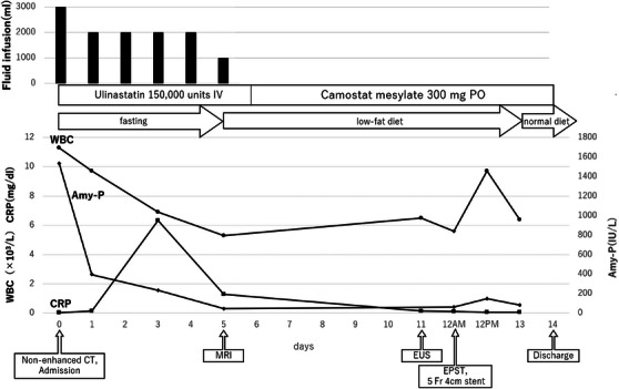
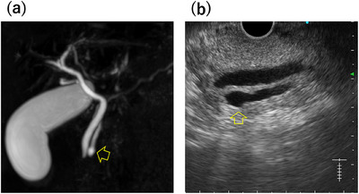
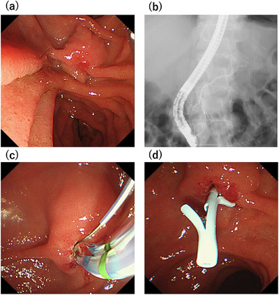
Wirsungocele, a cystic dilation at the end of the main pancreatic duct, is associated with recurrent acute pancreatitis. A 52-year-old man presented to our hospital with recurrent epigastric pain over an 8-month period with a history of multiple medical visits for the same complaint. Endoscopic ultrasound (EUS) and magnetic resonance cholangiopancreatography (MRCP) revealed focal cystic dilatation at the end of the main pancreatic duct; thus, he was diagnosed with Wirsungocele. He underwent endoscopic pancreatic sphincterotomy and 5Fr 4 cm pancreatic duct stent placement; the pancreatic duct stent was removed 1 month later. Magnetic resonance imaging performed 3 months after discharge revealed no cystic dilation, and he has had no recurrence of pancreatitis for at least 6 months. Dysfunction of the sphincter of Oddi, weakening of the pancreatic duct wall, inflammation and recurrent stress, elevated intraductal pressure, and genetic and structural factors are suspected mechanisms behind the pathophysiology of Wirsungocele. Although the etiology of Wirsungocele is not known, its timely identification and treatment are critical to preventing recurrent episodes of pancreatitis. This case demonstrates the diagnostic value of combining MRCP and EUS as well as the therapeutic benefits of endoscopic intervention, including sphincterotomy and stent placement, in managing Wirsungocele-associated recurrent pancreatitis. Given the paucity of reports on recurrent pancreatitis due to the Wirsungocele, we herein report this case and review the literature.
Keywords: endoscopic pancreatic sphincterotomy | magnetic resonance cholangiopancreatography | pancreatic duct stent | recurrent acute pancreatitis | wirsungocele.
© 2025 The Author(s). DEN Open published by John Wiley & Sons Australia, Ltd on behalf of Japan Gastroenterological Endoscopy Society.
Conflict of interest statement
The authors declare no conflicts of interest.
Figures
Similar articles
-
Treatment of Wirsungocele Induced Chronic Pancreatitis by Endoscopic Retrograde Cholangiopancreatography and Stent Placement: A Case Report.JGH Open. 2025 May 12;9(5):e70179. doi: 10.1002/jgh3.70179. eCollection 2025 May. JGH Open. 2025. PMID: 40365487 Free PMC article.
-
Recurrent acute pancreatitis and Wirsungocele. A case report and review of literature.JOP. 2008 Jul 10;9(4):531-3. JOP. 2008. PMID: 18648148 Review.
-
Wirsungocele: evaluation by MRCP and clinical significance.Abdom Radiol (NY). 2021 Feb;46(2):616-622. doi: 10.1007/s00261-020-02675-4. Epub 2020 Jul 31. Abdom Radiol (NY). 2021. PMID: 32737547 Free PMC article.
-
Value of Magnetic Resonance Cholangiopancreatography in Santorinicele and Wirsungocele.Curr Med Imaging. 2021;17(12):1451-1459. doi: 10.2174/1573405617666210804153921. Curr Med Imaging. 2021. PMID: 34348627
-
AGA Clinical Practice Update on the Endoscopic Approach to Recurrent Acute and Chronic Pancreatitis: Expert Review.Gastroenterology. 2022 Oct;163(4):1107-1114. doi: 10.1053/j.gastro.2022.07.079. Epub 2022 Aug 22. Gastroenterology. 2022. PMID: 36008176 Review.
References
-
- Abu‐Hamda E. M. and Baron T. H., “Cystic Dilatation of the Intraduodenal Portion of the Duct of Wirsung (Wirsungocele),” Gastrointestinal Endoscopy 59 (2004): 745–747. - PubMed
-
- Gupta R., Lakhtakia S., Tandan M., Santosh D., Rao G. V., and Reddy D. N., “Recurrent Acute Pancreatitis and Wirsungocele,” Journal of Pancreas 9 (2008): 531–533. - PubMed
-
- Zhao X. Z., Wan X., Luo M., et al., “Value of Magnetic Resonance Cholangiopancreatography in Santorinicele and Wirsungocele,” Current Medical Imaging 17 (2021): 1451–1459. - PubMed
LinkOut - more resources
Full Text Sources