. 2025 Jun;32(6):e70241.
doi: 10.1111/ene.70241.

Peripherin: A Novel Early Diagnostic and Prognostic Plasmatic Biomarker in Amyotrophic Lateral Sclerosis

Affiliations

Peripherin: A Novel Early Diagnostic and Prognostic Plasmatic Biomarker in Amyotrophic Lateral Sclerosis

Alessandro Bombaci et al. Eur J Neurol. 2025 Jun.

Abstract

Background: Motor neuron diseases (MND) are heterogeneous and complex neurodegenerative disorders. Biomarkers could facilitate early diagnosis, prognosis determination, and patient stratification. Among the most studied biomarkers are neurofilaments, with peripherin (PRPH), a specific type predominantly expressed in the peripheral nervous system, gaining attention. To date, no studies have evaluated PRPH in human plasma.

Methods: Sandwich-ELISA was used to quantify plasma peripherin from 120 MND (100 ALS, 4 PMA, 15 PLS), 73 MND-mimics, and 38 healthy-controls (HCs). Plasma was collected at diagnosis or some months earlier. 41 ALS were evaluated longitudinally. ALSFRSr, MRC, spirometry, genetic tests, disease progression rate (PR), blood examinations, and neuropsychological tests were performed. Statistical analyses included Kruskal-Wallis, Mann-Whitney, Cox regression, and Kaplan-Meier curves.

Results: Plasma PRPH levels differed significantly among groups (p < 0.0001), showing higher values in MND participants than MND mimics and HCs. Moreover, PRPH levels were elevated in PLS compared with HSP patients (p = 0.0001). Differences persisted after adjusting for age and sex. ROC curve demonstrated that PRPH discriminated MND from MND mimics (AUC = 0.85). Elevated PRPH correlated positively with ALSFRSr and lower motor neuron index, whereas inversely with disease progression rate. Higher PRPH levels at the beginning of the disease were associated with longer survival.

Discussion: Plasma PRPH is raised in MND, particularly ALS, from the earliest stages, distinguishing MND from mimics and correlating with clinical parameters and survival. This suggests PRPH may reflect an endogenous response of lower motor neuron to injury. Further multicenter studies are required to refine the diagnostic and prognostic utility of PRPH in MND.

Keywords: HSP; PLS; PRPH; fluid‐biomarker; mimics; neurodegeneration; neurofilament; regeneration.


Conflict of interest statement

The authors declare no conflicts of interest.

Figures

FIGURE 1
FIGURE 1
Levels of plasma PRPH in MND, MND‐mimics, and HCs. HCs, healthy controls; MND, motor neuron disease; PRPH, Peripherin; ****p ≤ 0.0001. Box plots show median and interquartile range.
FIGURE 2
FIGURE 2
Correlation between clinical parameters and PRPH in ALS patients. ALFRSr_4limb at T0, Ssum of item 4,5,6,7,8,9 of the ALSFRSr at baseline; ALSFRSr at T0, ALS Functional Rating Scale‐revised total score at baseline; ALSFRSr at T6, ALS Functional Rating Scale‐revised total score at month 6; ALSFRSr_noresp at T0, ALSFRSr without the respiratory item at baseline; ALSFRSr_noresp at T6, ALSFRSr without the respiratory item at month 6; FVC, Forced Vital Capacity; LMNI, Lower Motor Neuron Index; MiToS, Milano‐Torino staging system; PRPH, Peripherin.
FIGURE 3
FIGURE 3
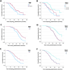
—Survival analysis. (A) and (B) represent the Kaplan–Meier analysis, respectively categorizing plasmatic PRPH levels using median and tertiles. (C) and (D) show survival curves covarying for plasmatic PRPH levels, sex, age at blood sampling, site of onset of the disease, and time from onset to death/tracheostomy, respectively categorizing plasmatic PRPH levels using median and tertiles. (E) and (F) show survival curves covarying for plasmatic PRPH levels, sex, age at blood sampling, site of onset of the disease, and time from blood sampling to death/tracheostomy, respectively categorizing plasmatic PRPH levels using median and tertiles. In (A), (C), and (E) the light‐blue line (status PRPH 0) corresponds to levels of PRPH below 1.35 ng/mL, whereas the red line (status PRPH 1) to levels of PRPH above 1.35 ng/mL; in (B), (D), and (E) the light‐blue line (status PRPH 0) corresponds to the 1° tertile (0.52–1.17 ng/mL), the red line (status PRPH 1) to levels of PRPH in the 2° tertile (1.19–1.72 ng/mL), and the light‐green line (status PRPH 2) to levels of PRPH in the 3° tertile (1.75–3.03 ng/mL). Patients with higher levels of PRPH show a longer survival.
FIGURE 4
FIGURE 4
Longitudinal changing of plasma PRPH. PRPH, Peripherin; ΔPRPH%, percentage of change in PRPH levels between T0 and T6.

References

    1. Hardiman O., Al‐Chalabi A., Chio A., et al., “Amyotrophic Lateral Sclerosis,” Nature Reviews Disease Primers 3, no. 1 (2017): 17071, 10.1038/nrdp.2017.71. - DOI - PubMed
    1. Chiò A., Calvo A., Moglia C., et al., “Phenotypic Heterogeneity of Amyotrophic Lateral Sclerosis: A Population Based Study,” Journal of Neurology, Neurosurgery, and Psychiatry 82, no. 7 (2011): 740–746, 10.1136/jnnp.2010.235952. - DOI - PubMed
    1. Renton A. E., Chiò A., and Traynor B. J., “State of Play in Amyotrophic Lateral Sclerosis Genetics,” Nature Neuroscience 17, no. 1 (2014): 17–23, 10.1038/nn.3584. - DOI - PMC - PubMed
    1. Steinacker P., Feneberg E., Weishaupt J., et al., “Neurofilaments in the Diagnosis of Motoneuron Diseases: A Prospective Study on 455 Patients,” Journal of Neurology, Neurosurgery, and Psychiatry 87, no. 1 (2016): 12–20, 10.1136/jnnp-2015-311387. - DOI - PubMed
    1. Lombardi V., Bombaci A., Zampedri L., et al., “Plasma pNfH Levels Differentiate SBMA From ALS,” Journal of Neurology, Neurosurgery, and Psychiatry 91 (2019): 215–217, 10.1136/jnnp-2019-320624. - DOI - PubMed