

Regional and Global Changes in Brain Structure 1-Year Post Pediatric "Mild" Traumatic Brain Injury
- PMID: 40476699
- PMCID: PMC12235941
- DOI: 10.1002/ana.27259
Regional and Global Changes in Brain Structure 1-Year Post Pediatric "Mild" Traumatic Brain Injury
Abstract
Objective: Despite the high prevalence of pediatric "mild" traumatic brain injury (pmTBI), very little is known about the long-term effects of injury on brain structure and how injuries manifest in the context of dynamic and regionally specific neurodevelopmental changes.
Methods: Prospective study design characterizing long-term effects of pmTBI on both global (brain age) and regional (hippocampal volume, hippocampal subfields, and cortical thickness) brain structure at approximately 7 days, 4 months, and 1-year post-injury. A large sample of age- and sex-matched healthy controls was imaged at identical temporal intervals to account for typical neurodevelopmental changes, and to assess how trauma potentially affects developmental trajectories.
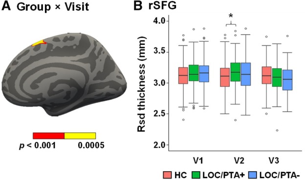
Results: A total of 269 pmTBI (age = 14.4 ± 2.9; 41.6% female) and 232 healthy controls (age = 14.1 ± 2.9; 44.8% female) were included in the final analyses (>70% 1-year retention). Results demonstrated that the presence of loss of consciousness and/or post-traumatic amnesia was associated with increased brain age up to 1-year post-injury, increased frontal cortical thickness at 4 months, as well as hippocampal atrophy across all time points relative to controls. "Mild" head trauma also interfered with hippocampal neurodevelopment in a dose-dependent fashion, including within the CA1 subfield. In contrast, post-concussive symptom burden was not associated with any structural abnormalities or alterations to neurodevelopment.
Interpretation: Current findings suggest a dose-dependent relationship between injury severity and alterations in both global and regional brain structure within the spectrum of pmTBI. Our results emphasize the importance of using objective biomarkers rather than subjective self-reported symptoms to better understand the long-term effects of injury. ANN NEUROL 2025;98:341-353.
© 2025 The Author(s). Annals of Neurology published by Wiley Periodicals LLC on behalf of American Neurological Association.
Conflict of interest statement
Nothing to report.
Figures


Similar articles
-
Dynamic Functional Connectivity in Pediatric Mild Traumatic Brain Injury.Neuroimage. 2024 Jan;285:120470. doi: 10.1016/j.neuroimage.2023.120470. Epub 2023 Nov 26. Neuroimage. 2024. PMID: 38016527 Free PMC article.
-
A one year longitudinal study of cortical myelination changes following pediatric mild traumatic brain injury.Neuroimage Clin. 2025 Jun 30;48:103837. doi: 10.1016/j.nicl.2025.103837. Online ahead of print. Neuroimage Clin. 2025. PMID: 40651061 Free PMC article.
-
Neural correlates of cognitive control deficits in pediatric mild traumatic brain injury.Hum Brain Mapp. 2023 Dec 1;44(17):6173-6184. doi: 10.1002/hbm.26504. Epub 2023 Oct 6. Hum Brain Mapp. 2023. PMID: 37800467 Free PMC article.
-
Psychological therapies for post-traumatic stress disorder and comorbid substance use disorder.Cochrane Database Syst Rev. 2016 Apr 4;4(4):CD010204. doi: 10.1002/14651858.CD010204.pub2. Cochrane Database Syst Rev. 2016. PMID: 27040448 Free PMC article.
-
Systemic pharmacological treatments for chronic plaque psoriasis: a network meta-analysis.Cochrane Database Syst Rev. 2017 Dec 22;12(12):CD011535. doi: 10.1002/14651858.CD011535.pub2. Cochrane Database Syst Rev. 2017. Update in: Cochrane Database Syst Rev. 2020 Jan 9;1:CD011535. doi: 10.1002/14651858.CD011535.pub3. PMID: 29271481 Free PMC article. Updated.
References
-
- Beauchamp MH, Ditchfield M, Maller JJ, et al. Hippocampus, amygdala and global brain changes 10 years after childhood traumatic brain injury. Int J Dev Neurosci 2011;29:137–143. - PubMed
MeSH terms
Grants and funding
- P30 GM122734/GM/NIGMS NIH HHS/United States
- R01 NS098494-03S1A1/NH/NIH HHS/United States
- 2R01NS098494-06A1/NH/NIH HHS/United States
- P30 GM122734/NH/NIH HHS/United States
- S10OD025313/NH/NIH HHS/United States
- R01 NS098494-01A1/NH/NIH HHS/United States
- S10 OD025313/OD/NIH HHS/United States
- R01 NS098494/NS/NINDS NIH HHS/United States
- P30 GM122734/NH/NIH HHS/United States
- R01 NS098494-01A1/NH/NIH HHS/United States
- R01 NS098494-03S1A1/NH/NIH HHS/United States
- 2R01NS098494-06A1/NH/NIH HHS/United States
- S10OD025313/NH/NIH HHS/United States
LinkOut - more resources
Full Text Sources
Medical
Miscellaneous
