. 2025 Jun;106(6):002120.
doi: 10.1099/jgv.0.002120.

Wisteria floribunda agglutinin enhances Zaire ebolavirus entry through interactions at specific N-linked glycosylation sites on the virus glycoprotein complex

Affiliations

Wisteria floribunda agglutinin enhances Zaire ebolavirus entry through interactions at specific N-linked glycosylation sites on the virus glycoprotein complex

Joshua D Duncan et al. J Gen Virol. 2025 Jun.

Abstract

Entry of Zaire ebolavirus (EBOV) into a host cell is a complex process requiring interactions between the viral glycoproteins (GPs) and cellular factors. These entry factors are cell-specific and can include cell surface lectins and phosphatidylserine receptors. Niemann-Pick type C1 is critical to the late stage of the entry process. Entry has been demonstrated to be enhanced by interactions between the virion and surface-expressed lectins, which interact with carbohydrate moieties attached to the GP. In addition, soluble lectins, including mannose-binding lectin, can enhance entry in vitro. However, the mechanism of lectin-mediated enhancement remains to be defined. This study investigated the possibility that plant lectins, Wisteria floribunda agglutinin (WFA), soybean agglutinin (SBA) and Galanthus nivalis agglutinin (GNA), which possess different carbohydrate-binding specificities, influence EBOV entry. WFA was observed to potently enhance entry of lentiviral pseudotype viruses (PVs) expressing the GP of three Ebolavirus species [Zaire, Sudan (Sudan ebolavirus) and Reston (Reston ebolavirus)], with the greatest impact on EBOV. SBA had a modest enhancing effect on entry that was specific to EBOV, whilst GNA had no impact on the entry of any of the Ebolavirus species. None of the lectins enhanced the entry of control PVs expressing the surface proteins of other RNA viruses tested. WFA was demonstrated to bind directly with the EBOV-GP via the glycans, and mutational analysis implicated N238 as contributing to the interaction. Furthermore, enhancement was observed in both human and bat cell lines, indicating a highly conserved mechanism of action. We conclude that the binding of WFA to EBOV-GP through interactions including the glycan at N238 results in GP alterations that enhance entry, providing evidence of a mechanism for lectin-mediated virus entry enhancement. Targeting lectin-ligand interactions presents a potential strategy for restricting Ebolavirus entry.

Keywords: Ebola virus; enhancement; lectin; pseudotype; virus entry.


Conflict of interest statement

The authors declare that there are no conflicts of interest.

Figures

Fig. 1.
Fig. 1.. Ebola virus GP-pseudotyped virus transduction is specifically enhanced in the presence of WFA. Pseudotyped virus particles possessing the envelope GPs of different viral species (including two strains of EBOV) were incubated with WFA, GNA or SBA prior to infection of human hepatoma (HuH7) cells. Each lectin was tested in a twofold dilution series in triplicate with a starting concentration of 20 µg ml−1. Data are reported as fold change in transduction relative to a no-lectin control group. EBOV-C15, Makona EBOV; EBOV-May, Mayinga EBOV. The y-axis differs between graphs to aid visualization. Statistical significance between the effect observed at the highest concentration and a no-lectin control was determined by a one-way ANOVA followed by Dunnett’s multiple comparison test, **P<0.01 and ****P<0.0001. Data are from a single representative experiment. Each data point represents the mean of three technical repeats with errors displaying sd.
Fig. 2.
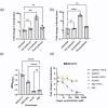
Fig. 2.. Direct interaction of WFA and EBOV-GP results in enhancement. WFA was added at a final concentration of 10 µg ml−1 to HuH7 cells at different stages of EBOV-C15 (a) and EBOV-May (b) PV entry. Cells were pre-treated with WFA prior to the addition of EBOV PVs. In the co-treatment condition, EBOV PVs and WFA were added simultaneously and incubated for 1 h. Post-treated WFA was added post-EBOV PV incubation. PV entry was measured by the luciferase reporter expression, and RLU values were compared with the untreated group. (c) The binding of pseudotypes to WFA was assessed by ELISA. Virus particles were incubated with coated WFA, and binding was revealed with incubation with anti-GP antibodies. Particles lacking GP were used as a negative control. (d) The specificity of interaction was determined by incubating GalNAc, GlcNAc or mannose with WFA during the co-treatment with EBOV-C15 pseudotype entry into HuH7 cells. Statistical significance was determined using one-way ANOVA followed by Dunnett’s multiple comparison test, P<0.01 (**), P<0.001 (***), P<0.0001 (****) and P>0.05 (ns). Data are from a single representative experiment. Each data point represents the mean of three technical repeats with error bars displaying sd.
Fig. 3.
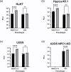
Fig. 3.. WFA-mediated enhancement of entry occurs in different species and is NPC1-dependent. Four mammalian cell lines, HuH7 (human, a) and HpyLu/45.1 (bat, b), human osteosarcoma cells (U2OS, c) and a validated U2OS NPC1-knock-out cell line (d), were transduced with pseudotypes possessing the GPs of Ebola C15 or VSV, +/−WFA at a concentration of 10 µg ml−1. Black bars indicate the control-treated pseudotypes, and grey bars indicate the WFA-treated pseudotypes. Significance was determined by one-way ANOVA followed by Sidak’s multiple comparison test, ns P>0.05 and ****P<0.0001. Data are from a single representative experiment. Each data point represents the mean of three technical repeats with error bars displaying sd.
Fig. 4.
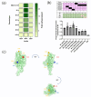
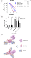
Fig. 4.. WFA-mediated enhancement of entry is modified in naturally occurring Ebolavirus variants. (a) Infection assays using HuH7 cells were performed using GPs from naturally occurring EBOV isolates with predicted altered N-linked glycosylation sites from the WT C15. Fold change between each lectin was compared to a no-lectin control. (b) Three in vitro-generated glycan mutants, M1, M2 and M3, were compared with variants B1, B12, B13, B14 and B16 in the enhancement assay at 10 µg ml−1. Differences were determined using one-way ANOVA and a Dunnett’s multiple comparison test, ns P>0.05, *P<0.05 and **P<0.01. (c) Representation of the sites of N-linked glycosylation on a model of the Mayinga EBOV-GP1 protein (produced using I-TASSER using the Makona C15 amino acid sequence modelled onto PDB structure 6VKM). Sites where removal of the glycan did not affect enhancement are highlighted in yellow, site (296) that increased enhancement is highlighted in blue and site (228) that reduced enhancement is highlighted in red.
Fig. 5.
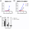
Fig. 5.. Enhanced entry of mucin domain-deleted mutant of Ebolavirus C15 by WFA. Transduction assays with HuH7 cells were performed in triplicate with EBOV-C15 (a) and EBOV-C15-ΔMLD (b) PVs using twofold serially diluted lectins starting from 10 µg ml−1. (c) Fold change in transduction of EBOV-C15 and EBOV-C15-ΔMLD at a lectin concentration of 10 µg ml−1. Black bars indicate pseudotypes possessing the EBOV-C15 GP; grey bars indicate pseudotypes possessing the EBOV-C15-ΔMLD GP. Comparisons were determined by one-way ANOVA followed by Sidak’s multiple comparison test, P>0.05 (ns) and P<0.0001 (****). Data are from a single representative experiment. Each data point represents the mean of three technical repeats with error bars displaying sd.
Fig. 6.
Fig. 6.. Modelling of WFA binding to EBOV-GP. (a) mAb KZ52 neutralization of EBOV-C15 pseudotype entry into HuH7 cells in the presence or absence of WFA lectin. EBOV PVs possessing the C15 or C15-ΔMLD GPs were incubated with a twofold serial dilution of KZ52 in the absence or presence of WFA, at a final concentration of 10 µg ml−1. Data were normalized to a no-antibody control, and each data point represents the mean of three technical repeats with error bars displaying sd. (b) Binding of KZ52 to EBOV PVs in a direct ELISA. Antibody was incubated with PVs in the presence or absence of WFA at 10 µg ml−1. Significance was determined by one-way ANOVA followed by Sidak’s multiple comparison test, ns P>0.05, ***P<0.001 and ****P<0.0001. Data are from a single representative experiment. Each data point represents the mean of three technical repeats with error bars displaying sd. (c) Modelling of the trimeric EBOV-GP in complex with KZ52 (PDB 3CSY) with one possible configuration of the tetrameric WFA molecule (PDB 5KXB) in close association with N228 of GP1 was performed using ChimeraX. Amino acid residues implicated in the NPC1 interaction are highlighted in red.

Similar articles

References

    1. Letafati A, Salahi Ardekani O, Karami H, Soleimani M. Ebola virus disease: a narrative review. Microb Pathog. 2023;181:106213. doi: 10.1016/j.micpath.2023.106213. - DOI - PubMed
    1. Feldmann H, Geisbert TW. Ebola haemorrhagic fever. Lancet. 2011;377:849–862. doi: 10.1016/S0140-6736(10)60667-8. - DOI - PMC - PubMed
    1. Aruna A, Mbala P, Minikulu L, Mukadi D, Bulemfu D, et al. Ebola virus disease outbreak - Democratic Republic of the Congo, August 2018-November 2019. MMWR Morb Mortal Wkly Rep. 2019;68:1162–1165. doi: 10.15585/mmwr.mm6850a3. - DOI - PMC - PubMed
    1. Kyobe Bosa H, Kamara N, Aragaw M, Wayengera M, Talisuna A, et al. The West Africa Ebola virus disease outbreak: 10 years on. Lancet Glob Heal. 2024;12:e1081–e1083. doi: 10.1016/S2214-109X(24)00129-3. - DOI - PubMed
    1. Feldmann H, Volchkov VE, Volchkova VA, Ströher U, Klenk H-D. Biosynthesis and role of filoviral glycoproteins. J Gen Virol. 2001;82:2839–2848. doi: 10.1099/0022-1317-82-12-2839. - DOI - PubMed