Review
. 2023;1(1):3.
doi: 10.1007/s44254-022-00002-2. Epub 2023 Mar 9.

Prone position: how understanding and clinical application of a technique progress with time

Affiliations
Review

Prone position: how understanding and clinical application of a technique progress with time

Luciano Gattinoni et al. Anesthesiol Perioper Sci. 2023.

Abstract

Historical background: The prone position was first proposed on theoretical background in 1974 (more advantageous distribution of mechanical ventilation). The first clinical report on 5 ARDS patients in 1976 showed remarkable improvement of oxygenation after pronation.

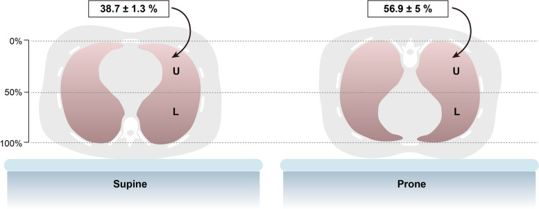
Pathophysiology: The findings in CT scans enhanced the use of prone position in ARDS patients. The main mechanism of the improved gas exchange seen in the prone position is nowadays attributed to a dorsal ventilatory recruitment, with a substantially unchanged distribution of perfusion. Regardless of the gas exchange, the primary effect of the prone position is a more homogenous distribution of ventilation, stress and strain, with similar size of pulmonary units in dorsal and ventral regions. In contrast, in the supine position the ventral regions are more expanded compared with the dorsal regions, which leads to greater ventral stress and strain, induced by mechanical ventilation.

Outcome in ards: The number of clinical studies paralleled the evolution of the pathophysiological understanding. The first two clinical trials in 2001 and 2004 were based on the hypothesis that better oxygenation would lead to a better survival and the studies were more focused on gas exchange than on lung mechanics. The equations better oxygenation = better survival was disproved by these and other larger trials (ARMA trial). However, the first studies provided signals that some survival advantages were possible in a more severe ARDS, where both oxygenation and lung mechanics were impaired. The PROSEVA trial finally showed the benefits of prone position on mortality supporting the thesis that the clinical advantages of prone position, instead of improved gas exchange, were mainly due to a less harmful mechanical ventilation and better distribution of stress and strain. In less severe ARDS, in spite of a better gas exchange, reduced mechanical stress and strain, and improved oxygenation, prone position was ineffective on outcome.

Prone position and covid-19: The mechanisms of oxygenation impairment in early COVID-19 are different than in typical ARDS and relate more on perfusion alteration than on alveolar consolidation/collapse, which are minimal in the early phase. Bronchial shunt may also contribute to the early COVID-19 hypoxemia. Therefore, in this phase, the oxygenation improvement in prone position is due to a better matching of local ventilation and perfusion, primarily caused by the perfusion component. Unfortunately, the conditions for improved outcomes, i.e. a better distribution of stress and strain, are almost absent in this phase of COVID-19 disease, as the lung parenchyma is nearly fully inflated. Due to some contradictory results, further studies are needed to better investigate the effect of prone position on outcome in COVID-19 patients.

Keywords: ARDS; Covid-19; Gas exchange; Lung mechanics; Prone position.


Conflict of interest statement

Competing interestsThe authors declare that they have no competing interests.

Figures

Fig. 1
Fig. 1
Number of studies per year about “prone position” and “ventilation”. The research was conducted in PubMed, suing as search query: “prone position” AND “ventilation”
Fig. 2
Fig. 2
Hydrostatic pressure – The sponge model. The lung of a patient with ARDS, characterized by interstitial edema, can be considered as a wet sponge. According to a gravity-dependent gradient, the edematous lung tissue exerts a hydrostatic pressure over the tissue below, leading to lung collapse in the dependent regions
Fig. 3
Fig. 3
CT-scans in supine vs. prone position at end-expiration. Representative CT-scans of a patient with ARDS, showing that lung densities redistribute from the dorsal to ventral regions when moving from supine to prone position
Fig. 4
Fig. 4
Chest wall compliance is the sum of three components: anterior (sternal), posterior (dorsal) and diaphragmatic. The anterior chest wall is more compliant than the posterior one, due to anatomical reasons. Therefore, in supine position the gas is mainly distributed primarily towards the anterior and diaphragmatic regions and less to the posterior ones. In prone position, the anterior chest wall compliance decreases, the dorsal one increases, though not reaching the values of the sternal compliance in supine. The diaphragmatic compliance is substantially unmodified. Thus, the final result is that in prone position the total respiratory system compliance decreases if the lung compliance remains unmodified
Fig. 5
Fig. 5
Homogenization of gas/tissue distribution. Representation of gas/tissue ratio, i.e. an index of regional lung inflation derived from CT-scans analysis, as a function of lung height in the supine (diamonds) and prone (circles) positions. A height of 0% refers to the nondependent surface of the thorax (ventral in the supine position and dorsal in the prone position). Conversely, a height of 100% refers to the dependent surface of the thorax. The green symbols refer to normal lungs (n = 14), while the red symbols to lungs of patients with ARDS (n = 20). Adapted from reference [32]
Fig. 6
Fig. 6
Thorax-lung shape mismatch. In this schematic representation of a transverse section of the thorax, the lung is represented as an orange triangle, while the pleural cavity is represented as a yellow oval. The isolated lung, in absence of gravity, can be thought of as a triangle, with all alveolar units of the same size. When the lung is placed into the thoracic cage, the apex of the cone stretches to adapt to the oval shape of the pleural cavity, which leads to an increase in size of the units in this area. When gravity is added, the units in the lower part of the lung tend to collapse due to the superimposed pressure of the units above. If the patient is then pronated, the hydrostatic pressure effect and the shape mismatch act in opposite directions, leading to a more homogeneous distribution of ventilation
Fig. 7
Fig. 7
Lung mass distribution. Due to its anatomical conformation, most of lung parenchyma is located in the dorsal part of the thorax. The letter “U” indicates the upper part of the lung, while the letter “L” indicates the lower one. At 50% of the sternum-vertebra distance, the nondependent lung mass in the supine position is less than 40%, while it rises to almost 60% in the prone position

Similar articles

Cited by

References

    1. Bryan AC. Conference on the scientific basis of respiratory therapy. Pulmonary physiotherapy in the pediatric age group. Comments of a devil’s advocate. Am Rev Respir Dis. 1974;110:143–4. - PubMed
    1. Piehl MA, Brown RS. Use of extreme position changes in acute respiratory failure. Crit Care Med. 1976;4:13–4. - PubMed
    1. Douglas WW, Rehder K, Beynen FM, Sessler AD, Marsh HM. Improved oxygenation in patients with acute respiratory failure: the prone position. Am Rev Respir Dis. 1977;115:559–66. - PubMed
    1. Maunder RJ, Shuman WP, McHugh JW, Marglin SI, Butler J. Preservation of normal lung regions in the adult respiratory distress syndrome. Analysis by computed tomography. JAMA. 1986;255:2463–5. - PubMed
    1. Gattinoni L, Mascheroni D, Torresin A, Marcolin R, Fumagalli R, Vesconi S, et al. Morphological response to positive end expiratory pressure in acute respiratory failure. Computerized tomography study. Intensive Care Med. 1986;12:137–42. - PubMed

LinkOut - more resources