

AI-guided spatiotemporal dispersion mapping for individualized ablation in an all-comer cohort with atrial fibrillation
- PMID: 40478367
- PMCID: PMC12476313
- DOI: 10.1007/s10840-025-02083-y
AI-guided spatiotemporal dispersion mapping for individualized ablation in an all-comer cohort with atrial fibrillation
Abstract
Background: Artificial intelligence (AI)-guided spatiotemporal dispersion (stD) mapping has been shown to improve outcomes in patients with persistent atrial fibrillation (AF). However, the relationship between stD mapping and markers of atrial cardiomyopathy, dispersion patterns in paroxysmal versus persistent AF, stability of dispersion regions, and stD-guided ablation-related outcomes in all-comer cohorts remain elusive.
Methods: In this retrospective single-center analysis, AF patients underwent high-density electroanatomical mapping alongside multiple instances of stD mapping using VOLTA AF Explorer software. Pulmonary vein isolation (PVI) and targeted ablation of left atrial dispersion regions were performed. Clinical, echocardiographic, biomarker, and low-voltage area (LVA) data were collected as markers of left atrial remodeling.
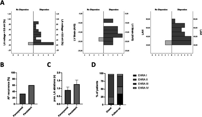
Results: stD mapping identified dispersion in 92% of patients. Mean time since AF diagnosis was 7 ± 1 years. Overall, 58% of patients showed dispersion exclusively co-localizing with low-voltage areas, while 42% had dispersion extending into intermediate or normal voltage regions. Dispersion burden correlated strongly with LVA extent and other remodeling markers such as NT-proBNP and LAVI. Persistent AF patients exhibited a significantly higher number of dispersion sites compared to paroxysmal AF. Dispersion patterns remained largely consistent before and after cardioversion in persistent AF, with the posterior left atrial wall emerging as a common hotspot. At follow-up, AF recurred in 33% of paroxysmal and 60% of persistent AF patients who had dispersion ablation limited to the left atrium. Despite these recurrences, most patients reported an improvement in symptomatic burden.
Conclusion: AI-guided stD mapping effectively identifies atrial remodeling beyond classical voltage-derived substrate, supporting its potential as a useful adjunctive tool in AF characterization.
Keywords: Artificial intelligence; Atrial fibrillation; Catheter ablation; Spatiotemporal dispersion; Substrate mapping.
© 2025. The Author(s).
Conflict of interest statement
Declarations. Competing interests: The authors declare that they have no competing interests relevant to the content of this article.
Figures


References
-
- Haissaguerre M, et al. Spontaneous initiation of atrial fibrillation by ectopic beats originating in the pulmonary veins. N Engl J Med. 1998;339(10):659–66. - PubMed
-
- Van Gelder, I.C., et al., 2024 ESC Guidelines for the management of atrial fibrillation developed in collaboration with the European Association for Cardio-Thoracic Surgery (EACTS): developed by the task force for the management of atrial fibrillation of the European Society of Cardiology (ESC), with the special contribution of the European Heart Rhythm Association (EHRA) of the ESC. Endorsed by the European Stroke Organisation (ESO). European Heart Journal, 2024. 45(36): p. 3314–3414. - PubMed
-
- Wong KCK, et al. No benefit of complex fractionated atrial electrogram ablation in addition to circumferential pulmonary vein ablation and linear ablation. Circ Arrhythm Electrophysiol. 2015;8(6):1316–24. - PubMed
-
- Takahashi Y, et al. Mapping-guided ablation for persistent atrial fibrillation (MAP-AF): a multicenter, single-blind, randomized controlled trial. Circ Arrhythm Electrophysiol. 2024;17(8):e012829. - PubMed
MeSH terms
LinkOut - more resources
Full Text Sources
Medical
Research Materials
